


 Foundational characteristics of cancer include proliferation, angiogenesis, migration, evasion of apoptosis, and cellular immortality. Find key markers for these cellular processes and antibodies to detect them.
 The SUMOplot™ Analysis Program predicts and scores sumoylation sites in your protein. SUMOylation is a post-translational modification involved in various cellular processes, such as nuclear-cytosolic transport, transcriptional regulation, apoptosis, protein stability, response to stress, and progression through the cell cycle.
 The Autophagy Receptor Motif Plotter predicts and scores autophagy receptor binding sites in your protein. Identifying proteins connected to this pathway is critical to understanding the role of autophagy in physiological as well as pathological processes such as development, differentiation, neurodegenerative diseases, stress, infection, and cancer.
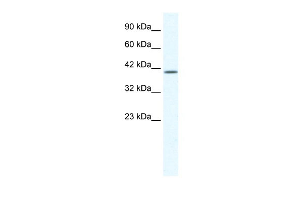
KCNK13 antibody - C-terminal region
Rabbit Polyclonal Antibody
- SPECIFICATION
- CITATIONS
- PROTOCOLS
- BACKGROUND

Application
| WB |
|---|---|
| Primary Accession | Q9HB14 |
| Other Accession | NM_022054, NP_071337 |
| Reactivity | Human, Horse, Bovine, Dog |
| Predicted | Human, Dog |
| Host | Rabbit |
| Clonality | Polyclonal |
| Calculated MW | 45kDa |
| Gene ID | 56659 |
|---|---|
| Alias Symbol | THIK1, THIK-1, K2p13.1 |
| Other Names | Potassium channel subfamily K member 13, Tandem pore domain halothane-inhibited potassium channel 1, THIK-1, KCNK13 |
| Format | Liquid. Purified antibody supplied in 1x PBS buffer with 0.09% (w/v) sodium azide and 2% sucrose. |
| Reconstitution & Storage | Add 100 ul of distilled water. Final anti-KCNK13 antibody concentration is 1 mg/ml in PBS buffer with 2% sucrose. For longer periods of storage, store at 20°C. Avoid repeat freeze-thaw cycles. |
| Precautions | KCNK13 antibody - C-terminal region is for research use only and not for use in diagnostic or therapeutic procedures. |
| Name | KCNK13 {ECO:0000303|PubMed:24163367, ECO:0000312|HGNC:HGNC:6275} |
|---|---|
| Function | K(+) channel that conducts outward rectifying tonic currents potentiated by purinergic signals (PubMed:24163367, PubMed:25148687, PubMed:30472253, PubMed:38409076). Homo- and heterodimerizes to form functional channels with distinct regulatory and gating properties (PubMed:25148687). Contributes most of K(+) currents at the plasma membrane of resting microglia. Maintains a depolarized membrane potential required for proper ramified microglia morphology and phagocytosis, selectively mediating microglial pruning of presynaptic compartments at hippocampal excitatory synapses (PubMed:38409076). Upon local release of ATP caused by neuronal injury or infection, it is potentiated by P2RY12 and P2RX7 receptor signaling and contributes to ATP-triggered K(+) efflux underlying microglial NLRP3 inflammasome assembly and IL1B release (By similarity) (PubMed:38409076). |
| Cellular Location | Cell membrane; Multi-pass membrane protein |
| Tissue Location | Expressed in microglia (at protein level). |

Thousands of laboratories across the world have published research that depended on the performance of antibodies from Abcepta to advance their research. Check out links to articles that cite our products in major peer-reviewed journals, organized by research category.
info@abcepta.com, and receive a free "I Love Antibodies" mug.

Provided below are standard protocols that you may find useful for product applications.
References
Rajan,S.,etal.,(2001)J.Biol.Chem.276(10),7302-7311ReconstitutionandStorage:Forshorttermuse,storeat2-8Cupto1week.Forlongtermstorage,storeat-20Cinsmallaliquotstopreventfreeze-thawcycles.
If you have used an Abcepta product and would like to share how it has performed, please click on the "Submit Review" button and provide the requested information. Our staff will examine and post your review and contact you if needed.
If you have any additional inquiries please email technical services at tech@abcepta.com.
Ordering Information
Other Products
Shipping Information
