


 Foundational characteristics of cancer include proliferation, angiogenesis, migration, evasion of apoptosis, and cellular immortality. Find key markers for these cellular processes and antibodies to detect them.
 The SUMOplot™ Analysis Program predicts and scores sumoylation sites in your protein. SUMOylation is a post-translational modification involved in various cellular processes, such as nuclear-cytosolic transport, transcriptional regulation, apoptosis, protein stability, response to stress, and progression through the cell cycle.
 The Autophagy Receptor Motif Plotter predicts and scores autophagy receptor binding sites in your protein. Identifying proteins connected to this pathway is critical to understanding the role of autophagy in physiological as well as pathological processes such as development, differentiation, neurodegenerative diseases, stress, infection, and cancer.
> home > Products > Primary Antibodies > Antibody Collections > Inflammatory response > KNG1 antibody - middle region
KNG1 antibody - middle region
Rabbit Polyclonal Antibody
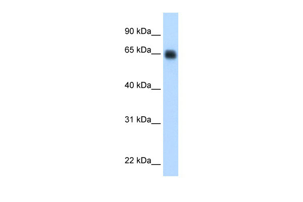
- SPECIFICATION
- CITATIONS
- PROTOCOLS
- BACKGROUND

Application
| WB |
|---|---|
| Primary Accession | P01042 |
| Other Accession | NM_001102416, NP_001095886 |
| Reactivity | Human, Mouse, Pig, Horse |
| Predicted | Human, Mouse, Pig, Horse |
| Host | Rabbit |
| Clonality | Polyclonal |
| Calculated MW | 72kDa |
| Gene ID | 3827 |
|---|---|
| Alias Symbol | BDK, KNG |
| Other Names | Kininogen-1, Alpha-2-thiol proteinase inhibitor, Fitzgerald factor, High molecular weight kininogen, HMWK, Williams-Fitzgerald-Flaujeac factor, Kininogen-1 heavy chain, T-kinin, Ile-Ser-Bradykinin, Bradykinin, Kallidin I, Lysyl-bradykinin, Kallidin II, Kininogen-1 light chain, Low molecular weight growth-promoting factor, KNG1, BDK, KNG |
| Format | Liquid. Purified antibody supplied in 1x PBS buffer with 0.09% (w/v) sodium azide and 2% sucrose. |
| Reconstitution & Storage | Add 50 ul of distilled water. Final anti-KNG1 antibody concentration is 1 mg/ml in PBS buffer with 2% sucrose. For longer periods of storage, store at 20°C. Avoid repeat freeze-thaw cycles. |
| Precautions | KNG1 antibody - middle region is for research use only and not for use in diagnostic or therapeutic procedures. |
| Name | KNG1 |
|---|---|
| Synonyms | BDK, KNG |
| Function | Kininogens are inhibitors of thiol proteases. HMW-kininogen plays an important role in blood coagulation by helping to position optimally prekallikrein and factor XI next to factor XII; HMW-kininogen inhibits the thrombin- and plasmin-induced aggregation of thrombocytes. LMW-kininogen inhibits the aggregation of thrombocytes. LMW-kininogen is in contrast to HMW-kininogen not involved in blood clotting. |
| Cellular Location | Secreted, extracellular space. |
| Tissue Location | Secreted in plasma. T-kinin is detected in malignant ovarian, colon and breast carcinomas, but not in benign tumors. |
Research Areas
Citations (0)

Thousands of laboratories across the world have published research that depended on the performance of antibodies from Abcepta to advance their research. Check out links to articles that cite our products in major peer-reviewed journals, organized by research category.
Submit your citation using an Abcepta antibody to
info@abcepta.com, and receive a free "I Love Antibodies" mug.
info@abcepta.com, and receive a free "I Love Antibodies" mug.

Application Protocols
Provided below are standard protocols that you may find useful for product applications.
Abcepta welcomes feedback from its customers.
If you have used an Abcepta product and would like to share how it has performed, please click on the "Submit Review" button and provide the requested information. Our staff will examine and post your review and contact you if needed.
If you have any additional inquiries please email technical services at tech@abcepta.com.
$ 389.00
Cat# AI12604
Ordering Information
United States
AlbaniaAustraliaAustriaBelgiumBosnia & HerzegovinaBrazilBulgariaCanadaCentral AmericaChinaCroatiaCyprusCzech RepublicDenmarkEstoniaFinlandFranceGermanyGreeceHong KongHungaryIcelandIndiaIndonesiaIrelandIsraelItalyJapanLatviaLithuaniaLuxembourgMacedoniaMalaysiaMaltaMexicoNetherlandsNew ZealandNorwayPakistanPolandPortugalRomaniaSerbiaSingaporeSlovakiaSloveniaSouth AfricaSouth KoreaSpainSwedenSwitzerlandTaiwanTurkeyUnited KingdomUnited StatesVietnamWorldwideOthers
USA Headquarters
(888) 735-7227 / (858) 622-0099 or (858) 875-1900
Other Products
Shipping Information
Domestic orders (in stock items)
Shipped out the same day. Orders placed after 1 PM (PST) will ship out the next business day.
International orders
Contact your local distributors
