


 Foundational characteristics of cancer include proliferation, angiogenesis, migration, evasion of apoptosis, and cellular immortality. Find key markers for these cellular processes and antibodies to detect them.
 The SUMOplot™ Analysis Program predicts and scores sumoylation sites in your protein. SUMOylation is a post-translational modification involved in various cellular processes, such as nuclear-cytosolic transport, transcriptional regulation, apoptosis, protein stability, response to stress, and progression through the cell cycle.
 The Autophagy Receptor Motif Plotter predicts and scores autophagy receptor binding sites in your protein. Identifying proteins connected to this pathway is critical to understanding the role of autophagy in physiological as well as pathological processes such as development, differentiation, neurodegenerative diseases, stress, infection, and cancer.
IL11RA antibody - middle region
Rabbit Polyclonal Antibody
- SPECIFICATION
- CITATIONS
- PROTOCOLS
- BACKGROUND

Application
| WB |
|---|---|
| Primary Accession | Q14626 |
| Other Accession | NM_004512, NP_004503 |
| Reactivity | Human, Mouse, Rat, Rabbit, Horse, Bovine, Guinea Pig, Dog |
| Predicted | Human, Mouse, Rabbit, Horse, Bovine, Guinea Pig, Dog |
| Host | Rabbit |
| Clonality | Polyclonal |
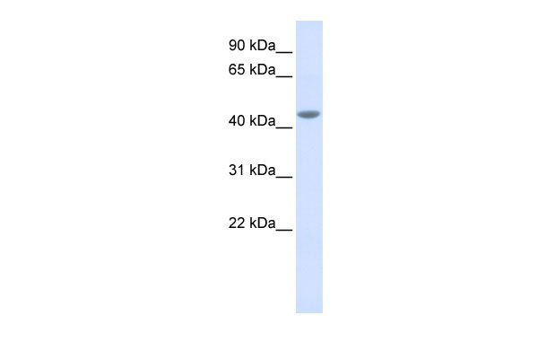
| Calculated MW | 43kDa |
| Gene ID | 3590 |
|---|---|
| Alias Symbol | MGC2146, CRSDA |
| Other Names | Interleukin-11 receptor subunit alpha, IL-11 receptor subunit alpha, IL-11R subunit alpha, IL-11R-alpha, IL-11RA, IL11RA |
| Format | Liquid. Purified antibody supplied in 1x PBS buffer with 0.09% (w/v) sodium azide and 2% sucrose. |
| Reconstitution & Storage | Add 50 ul of distilled water. Final anti-IL11RA antibody concentration is 1 mg/ml in PBS buffer with 2% sucrose. For longer periods of storage, store at 20°C. Avoid repeat freeze-thaw cycles. |
| Precautions | IL11RA antibody - middle region is for research use only and not for use in diagnostic or therapeutic procedures. |
| Name | IL11RA (HGNC:5967) |
|---|---|
| Function | Receptor for interleukin-11 (IL11). The receptor systems for IL6, LIF, OSM, CNTF, IL11 and CT1 can utilize IL6ST for initiating signal transmission. The IL11/IL11RA/IL6ST complex may be involved in the control of proliferation and/or differentiation of skeletogenic progenitor or other mesenchymal cells (Probable). Essential for the normal development of craniofacial bones and teeth. Restricts suture fusion and tooth number. |
| Cellular Location | [Interleukin-11 receptor subunit alpha]: Membrane; Single-pass type I membrane protein [Isoform HCR2]: Secreted |
| Tissue Location | Expressed in a number of cell lines, including the myelogenous leukemia cell line K-562, the megakaryocytic leukemia cell line M-07e, the erythroleukemia cell line TF-1, and the osteosarcoma cell lines, MG-63 and SaOS-2 (PubMed:7670098). Also expressed in normal and malignant prostate epithelial cell lines. Expression levels are increased in prostate carcinoma. |

Thousands of laboratories across the world have published research that depended on the performance of antibodies from Abcepta to advance their research. Check out links to articles that cite our products in major peer-reviewed journals, organized by research category.
info@abcepta.com, and receive a free "I Love Antibodies" mug.

Provided below are standard protocols that you may find useful for product applications.
References
Nakayama,T.,(2007)Int.J.Oncol.30(4),825-833ReconstitutionandStorage:Forshorttermuse,storeat2-8Cupto1week.Forlongtermstorage,storeat-20Cinsmallaliquotstopreventfreeze-thawcycles.
If you have used an Abcepta product and would like to share how it has performed, please click on the "Submit Review" button and provide the requested information. Our staff will examine and post your review and contact you if needed.
If you have any additional inquiries please email technical services at tech@abcepta.com.
Ordering Information
Other Products
Shipping Information
