


 Foundational characteristics of cancer include proliferation, angiogenesis, migration, evasion of apoptosis, and cellular immortality. Find key markers for these cellular processes and antibodies to detect them.
 The SUMOplot™ Analysis Program predicts and scores sumoylation sites in your protein. SUMOylation is a post-translational modification involved in various cellular processes, such as nuclear-cytosolic transport, transcriptional regulation, apoptosis, protein stability, response to stress, and progression through the cell cycle.
 The Autophagy Receptor Motif Plotter predicts and scores autophagy receptor binding sites in your protein. Identifying proteins connected to this pathway is critical to understanding the role of autophagy in physiological as well as pathological processes such as development, differentiation, neurodegenerative diseases, stress, infection, and cancer.
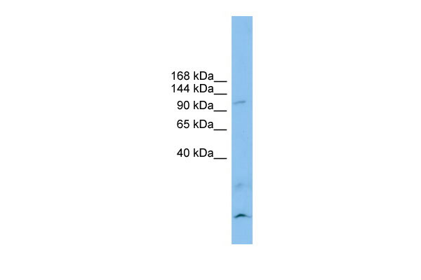
CLSTN3 antibody - N-terminal region
Rabbit Polyclonal Antibody
- SPECIFICATION
- CITATIONS
- PROTOCOLS
- BACKGROUND

Application
| WB |
|---|---|
| Primary Accession | Q9BQT9 |
| Other Accession | NM_014718, NP_055533 |
| Reactivity | Human, Mouse, Rat, Rabbit, Pig, Horse, Bovine, Guinea Pig, Dog |
| Predicted | Human, Mouse, Rat, Chicken, Guinea Pig |
| Host | Rabbit |
| Clonality | Polyclonal |
| Calculated MW | 106kDa |
| Gene ID | 9746 |
|---|---|
| Alias Symbol | CSTN3, KIAA0726, MGC131797, MGC138488, alcbeta, CDHR14 |
| Other Names | Calsyntenin-3, Alcadein-beta, Alc-beta, CLSTN3, CS3, KIAA0726 |
| Format | Liquid. Purified antibody supplied in 1x PBS buffer with 0.09% (w/v) sodium azide and 2% sucrose. |
| Reconstitution & Storage | Add 50 ul of distilled water. Final anti-CLSTN3 antibody concentration is 1 mg/ml in PBS buffer with 2% sucrose. For longer periods of storage, store at 20°C. Avoid repeat freeze-thaw cycles. |
| Precautions | CLSTN3 antibody - N-terminal region is for research use only and not for use in diagnostic or therapeutic procedures. |
| Name | CLSTN3 {ECO:0000303|PubMed:33101212, ECO:0000312|HGNC:HGNC:18371} |
|---|---|
| Function | Postsynaptic adhesion molecule that binds to presynaptic neurexins to mediate both excitatory and inhibitory synapse formation (PubMed:25352602). Promotes synapse development by acting as a cell adhesion molecule at the postsynaptic membrane, which associates with both neurexin-alpha and neurexin-beta proteins at the presynaptic membrane (PubMed:25352602). Regulates the balance between excitatory and inhibitory synapses by inhibiting formation of excitatory parallel- fiber synapses and promoting formation of inhibitory synapses in the same neuron (By similarity). May also be involved in ascorbate (vitamin C) uptake via its interaction with SLC23A2/SVCT2 (PubMed:34673103). Complex formation with APBA2 and APP, stabilizes APP metabolism and enhances APBA2-mediated suppression of beta-APP40 secretion, due to the retardation of intracellular APP maturation (Probable) (PubMed:12972431). |
| Cellular Location | Postsynaptic cell membrane {ECO:0000250|UniProtKB:Q99JH7}; Single-pass type I membrane protein. Endoplasmic reticulum membrane {ECO:0000250|UniProtKB:Q99JH7}; Single-pass type I membrane protein. Golgi apparatus membrane {ECO:0000250|UniProtKB:Q99JH7}; Single-pass type I membrane protein. Cell projection, dendrite {ECO:0000250|UniProtKB:Q99JH7}. Note=Most prominent in the postsynaptic specializations of asymmetric (type I) synapses with both axodendritic and axospinous localization {ECO:0000250|UniProtKB:Q99JH7} |
| Tissue Location | According to PubMed:12498782, expressed predominantly in the brain and in kidney (PubMed:12498782). Low levels in heart, skeletal muscle, liver, placenta, pancreas and lung (PubMed:12498782). According to PubMed:12972431, predominant expression in brain, and only marginal in kidney (PubMed:12972431). In brain, present throughout all cortical layers, highest levels in GABAergic neurons (based on morphology and distribution pattern) (PubMed:12972431). |
Citations (0)

Thousands of laboratories across the world have published research that depended on the performance of antibodies from Abcepta to advance their research. Check out links to articles that cite our products in major peer-reviewed journals, organized by research category.
Submit your citation using an Abcepta antibody to
info@abcepta.com, and receive a free "I Love Antibodies" mug.
info@abcepta.com, and receive a free "I Love Antibodies" mug.

Application Protocols
Provided below are standard protocols that you may find useful for product applications.
Abcepta welcomes feedback from its customers.
If you have used an Abcepta product and would like to share how it has performed, please click on the "Submit Review" button and provide the requested information. Our staff will examine and post your review and contact you if needed.
If you have any additional inquiries please email technical services at tech@abcepta.com.
$ 389.00
Cat# AI12745
Ordering Information
United States
AlbaniaAustraliaAustriaBelgiumBosnia & HerzegovinaBrazilBulgariaCanadaCentral AmericaChinaCroatiaCyprusCzech RepublicDenmarkEstoniaFinlandFranceGermanyGreeceHong KongHungaryIcelandIndiaIndonesiaIrelandIsraelItalyJapanLatviaLithuaniaLuxembourgMacedoniaMalaysiaMaltaMexicoNetherlandsNew ZealandNorwayPakistanPolandPortugalRomaniaSerbiaSingaporeSlovakiaSloveniaSouth AfricaSouth KoreaSpainSwedenSwitzerlandTaiwanTurkeyUnited KingdomUnited StatesVietnamWorldwideOthers
USA Headquarters
(888) 735-7227 / (858) 622-0099 or (858) 875-1900
Other Products
Shipping Information
Domestic orders (in stock items)
Shipped out the same day. Orders placed after 1 PM (PST) will ship out the next business day.
International orders
Contact your local distributors
