


 Foundational characteristics of cancer include proliferation, angiogenesis, migration, evasion of apoptosis, and cellular immortality. Find key markers for these cellular processes and antibodies to detect them.
 The SUMOplot™ Analysis Program predicts and scores sumoylation sites in your protein. SUMOylation is a post-translational modification involved in various cellular processes, such as nuclear-cytosolic transport, transcriptional regulation, apoptosis, protein stability, response to stress, and progression through the cell cycle.
 The Autophagy Receptor Motif Plotter predicts and scores autophagy receptor binding sites in your protein. Identifying proteins connected to this pathway is critical to understanding the role of autophagy in physiological as well as pathological processes such as development, differentiation, neurodegenerative diseases, stress, infection, and cancer.
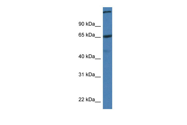
St6galnac1 antibody - C-terminal region
Rabbit Polyclonal Antibody
- SPECIFICATION
- CITATIONS
- PROTOCOLS
- BACKGROUND

Application
| WB |
|---|---|
| Primary Accession | Q9QZ39 |
| Other Accession | NM_011371, NP_035501 |
| Reactivity | Human, Mouse, Rat, Rabbit, Pig, Horse, Bovine, Guinea Pig, Dog |
| Predicted | Mouse, Rat, Rabbit, Pig, Horse, Bovine, Dog |
| Host | Rabbit |
| Clonality | Polyclonal |
| Calculated MW | 61kDa |
| Gene ID | 20445 |
|---|---|
| Alias Symbol | Siat7a |
| Other Names | Alpha-N-acetylgalactosaminide alpha-2, 6-sialyltransferase 1, 2.4.99.3, GalNAc alpha-2, 6-sialyltransferase I, ST6GalNAc I, ST6GalNAcI, Sialyltransferase 7A, SIAT7-A, St6galnac1, Siat7a |
| Format | Liquid. Purified antibody supplied in 1x PBS buffer with 0.09% (w/v) sodium azide and 2% sucrose. |
| Reconstitution & Storage | Add 50 ul of distilled water. Final anti-St6galnac1 antibody concentration is 1 mg/ml in PBS buffer with 2% sucrose. For longer periods of storage, store at 20°C. Avoid repeat freeze-thaw cycles. |
| Precautions | St6galnac1 antibody - C-terminal region is for research use only and not for use in diagnostic or therapeutic procedures. |
| Name | St6galnac1 {ECO:0000312|MGI:MGI:1341826} |
|---|---|
| Synonyms | Siat7a |
| Function | Protein sialyltransferase specifically expressed in goblet cells that plays a key role in intestinal host-commensal homeostasis (PubMed:35303419). Conjugates sialic acid with an alpha-2-6 linkage to N-acetylgalactosamine (GalNAc) glycan chains linked to serine or threonine in glycoproteins (PubMed:10788794). Catalyzes the formation of the sialyl-Tn (S-Tn) antigen, an antigen found in intestinal goblet cells (PubMed:35303419, PubMed:16207894). Protein sialylation in globlet cells is essential for mucus integrity and is required to protect the intestinal mucus against excessive bacterial proteolytic degradation (PubMed:35303419). |
| Cellular Location | Golgi apparatus membrane {ECO:0000250|UniProtKB:Q9NSC7}; Single-pass type II membrane protein {ECO:0000250|UniProtKB:Q9NSC7} |
| Tissue Location | Submaxillary gland, mammary gland, spleen and colon. |
Research Areas
Citations (0)

Thousands of laboratories across the world have published research that depended on the performance of antibodies from Abcepta to advance their research. Check out links to articles that cite our products in major peer-reviewed journals, organized by research category.
Submit your citation using an Abcepta antibody to
info@abcepta.com, and receive a free "I Love Antibodies" mug.
info@abcepta.com, and receive a free "I Love Antibodies" mug.

Application Protocols
Provided below are standard protocols that you may find useful for product applications.
Abcepta welcomes feedback from its customers.
If you have used an Abcepta product and would like to share how it has performed, please click on the "Submit Review" button and provide the requested information. Our staff will examine and post your review and contact you if needed.
If you have any additional inquiries please email technical services at tech@abcepta.com.
$ 389.00
Cat# AI12798
Ordering Information
United States
AlbaniaAustraliaAustriaBelgiumBosnia & HerzegovinaBrazilBulgariaCanadaCentral AmericaChinaCroatiaCyprusCzech RepublicDenmarkEstoniaFinlandFranceGermanyGreeceHong KongHungaryIcelandIndiaIndonesiaIrelandIsraelItalyJapanLatviaLithuaniaLuxembourgMacedoniaMalaysiaMaltaMexicoNetherlandsNew ZealandNorwayPakistanPolandPortugalRomaniaSerbiaSingaporeSlovakiaSloveniaSouth AfricaSouth KoreaSpainSwedenSwitzerlandTaiwanTurkeyUnited KingdomUnited StatesVietnamWorldwideOthers
USA Headquarters
(888) 735-7227 / (858) 622-0099 or (858) 875-1900
Other Products
Shipping Information
Domestic orders (in stock items)
Shipped out the same day. Orders placed after 1 PM (PST) will ship out the next business day.
International orders
Contact your local distributors
