


 Foundational characteristics of cancer include proliferation, angiogenesis, migration, evasion of apoptosis, and cellular immortality. Find key markers for these cellular processes and antibodies to detect them.
 The SUMOplot™ Analysis Program predicts and scores sumoylation sites in your protein. SUMOylation is a post-translational modification involved in various cellular processes, such as nuclear-cytosolic transport, transcriptional regulation, apoptosis, protein stability, response to stress, and progression through the cell cycle.
 The Autophagy Receptor Motif Plotter predicts and scores autophagy receptor binding sites in your protein. Identifying proteins connected to this pathway is critical to understanding the role of autophagy in physiological as well as pathological processes such as development, differentiation, neurodegenerative diseases, stress, infection, and cancer.
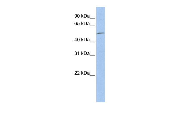
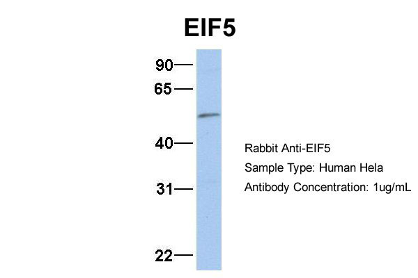
EIF5 antibody - N-terminal region
Rabbit Polyclonal Antibody
- SPECIFICATION
- CITATIONS
- PROTOCOLS
- BACKGROUND

Application
| WB |
|---|---|
| Primary Accession | P55010 |
| Other Accession | NM_183004, NP_892116 |
| Reactivity | Human, Mouse, Rat, Rabbit, Horse, Bovine, Guinea Pig, Dog |
| Predicted | Human, Mouse, Rat, Chicken, Horse, Bovine, Dog |
| Host | Rabbit |
| Clonality | Polyclonal |
| Calculated MW | 49kDa |
| Gene ID | 1983 |
|---|---|
| Alias Symbol | EIF-5A |
| Other Names | Eukaryotic translation initiation factor 5, eIF-5, EIF5 |
| Format | Liquid. Purified antibody supplied in 1x PBS buffer with 0.09% (w/v) sodium azide and 2% sucrose. |
| Reconstitution & Storage | Add 50 ul of distilled water. Final anti-EIF5 antibody concentration is 1 mg/ml in PBS buffer with 2% sucrose. For longer periods of storage, store at 20°C. Avoid repeat freeze-thaw cycles. |
| Precautions | EIF5 antibody - N-terminal region is for research use only and not for use in diagnostic or therapeutic procedures. |
| Name | EIF5 |
|---|---|
| Function | Component of the 43S pre-initiation complex (43S PIC), which binds to the mRNA cap-proximal region, scans mRNA 5'-untranslated region, and locates the initiation codon (PubMed:11166181, PubMed:22813744, PubMed:24319994). In this complex, acts as a GTPase- activating protein, by promoting GTP hydrolysis by eIF2G (EIF2S3) (PubMed:11166181). During scanning, interacts with both EIF1 (via its C-terminal domain (CTD)) and EIF1A (via its NTD) (PubMed:22813744). This interaction with EIF1A contributes to the maintenance of EIF1 within the open 43S PIC (PubMed:24319994). When start codon is recognized, EIF5, via its NTD, induces eIF2G (EIF2S3) to hydrolyze the GTP (PubMed:11166181). Start codon recognition also induces a conformational change of the PIC to a closed state (PubMed:22813744). This change increases the affinity of EIF5-CTD for EIF2-beta (EIF2S2), which allows the release, by an indirect mechanism, of EIF1 from the PIC (PubMed:22813744). Finally, EIF5 stabilizes the PIC in its closed conformation (PubMed:22813744). |
| Cellular Location | Cytoplasm. |

Thousands of laboratories across the world have published research that depended on the performance of antibodies from Abcepta to advance their research. Check out links to articles that cite our products in major peer-reviewed journals, organized by research category.
info@abcepta.com, and receive a free "I Love Antibodies" mug.

Provided below are standard protocols that you may find useful for product applications.
References
Si K.,et al.J. Biol. Chem. 271:16934-16938(1996).
Wiemann S.,et al.Genome Res. 11:422-435(2001).
Ota T.,et al.Nat. Genet. 36:40-45(2004).
Bechtel S.,et al.BMC Genomics 8:399-399(2007).
Mural R.J.,et al.Submitted (JUL-2005) to the EMBL/GenBank/DDBJ databases.
If you have used an Abcepta product and would like to share how it has performed, please click on the "Submit Review" button and provide the requested information. Our staff will examine and post your review and contact you if needed.
If you have any additional inquiries please email technical services at tech@abcepta.com.
Ordering Information
Other Products
Shipping Information
