


 Foundational characteristics of cancer include proliferation, angiogenesis, migration, evasion of apoptosis, and cellular immortality. Find key markers for these cellular processes and antibodies to detect them.
 The SUMOplot™ Analysis Program predicts and scores sumoylation sites in your protein. SUMOylation is a post-translational modification involved in various cellular processes, such as nuclear-cytosolic transport, transcriptional regulation, apoptosis, protein stability, response to stress, and progression through the cell cycle.
 The Autophagy Receptor Motif Plotter predicts and scores autophagy receptor binding sites in your protein. Identifying proteins connected to this pathway is critical to understanding the role of autophagy in physiological as well as pathological processes such as development, differentiation, neurodegenerative diseases, stress, infection, and cancer.
RP2 antibody - middle region
Rabbit Polyclonal Antibody
- SPECIFICATION
- CITATIONS
- PROTOCOLS
- BACKGROUND

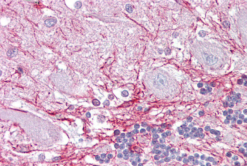
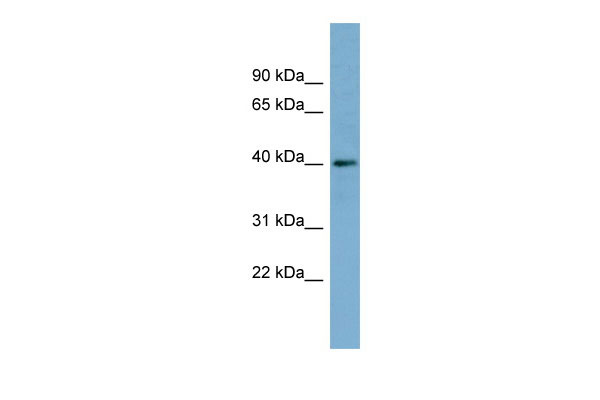
Application
| WB, IHC |
|---|---|
| Primary Accession | O75695 |
| Other Accession | NM_006003, NP_008846 |
| Reactivity | Human, Mouse, Rat, Rabbit, Horse, Bovine, Guinea Pig, Dog |
| Predicted | Human, Mouse, Rabbit, Horse, Bovine, Dog |
| Host | Rabbit |
| Clonality | Polyclonal |
| Calculated MW | 40kDa |
| Gene ID | 6102 |
|---|---|
| Alias Symbol | KIAA0215, TBCCD2, XRP2, NME10, DELXp11.3 |
| Other Names | Protein XRP2, RP2 |
| Format | Liquid. Purified antibody supplied in 1x PBS buffer with 0.09% (w/v) sodium azide and 2% sucrose. |
| Reconstitution & Storage | Add 50 ul of distilled water. Final anti-RP2 antibody concentration is 1 mg/ml in PBS buffer with 2% sucrose. For longer periods of storage, store at 20°C. Avoid repeat freeze-thaw cycles. |
| Precautions | RP2 antibody - middle region is for research use only and not for use in diagnostic or therapeutic procedures. |
| Name | RP2 |
|---|---|
| Function | Acts as a GTPase-activating protein (GAP) involved in trafficking between the Golgi and the ciliary membrane. Involved in localization of proteins, such as NPHP3, to the cilium membrane by inducing hydrolysis of GTP ARL3, leading to the release of UNC119 (or UNC119B). Acts as a GTPase-activating protein (GAP) for tubulin in concert with tubulin-specific chaperone C, but does not enhance tubulin heterodimerization. Acts as a guanine nucleotide dissociation inhibitor towards ADP-ribosylation factor-like proteins. |
| Cellular Location | Cell membrane; Lipid-anchor; Cytoplasmic side. Cell projection, cilium. Note=Detected predominantly at the plasma membrane of rod and cone photoreceptors. Not detected in the nucleus. |
| Tissue Location | Ubiquitous. Expressed in the rod and cone photoreceptors, extending from the tips of the outer segment (OS) through the inner segment (IS) and outer nuclear layer (ONL) and into the synaptic terminals of the outer plexiform layer (ONL). Also detected in the bipolar, horizontal and amacrine cells in the inner nuclear layer (INL), extending to the inner plexiform layer (IPL) and though the ganglion cell layer (GCL) and into the nerve fiber layer (NFL) (at protein level). |

Thousands of laboratories across the world have published research that depended on the performance of antibodies from Abcepta to advance their research. Check out links to articles that cite our products in major peer-reviewed journals, organized by research category.
info@abcepta.com, and receive a free "I Love Antibodies" mug.

Provided below are standard protocols that you may find useful for product applications.
References
Schwahn U.,et al.Nat. Genet. 19:327-332(1998).
Ross M.T.,et al.Nature 434:325-337(2005).
Burkard T.R.,et al.BMC Syst. Biol. 5:17-17(2011).
Chapple J.P.,et al.Hum. Mol. Genet. 9:1919-1926(2000).
Bartolini F.,et al.J. Biol. Chem. 277:14629-14634(2002).
If you have used an Abcepta product and would like to share how it has performed, please click on the "Submit Review" button and provide the requested information. Our staff will examine and post your review and contact you if needed.
If you have any additional inquiries please email technical services at tech@abcepta.com.
Ordering Information
Other Products
Shipping Information
