


 Foundational characteristics of cancer include proliferation, angiogenesis, migration, evasion of apoptosis, and cellular immortality. Find key markers for these cellular processes and antibodies to detect them.
 The SUMOplot™ Analysis Program predicts and scores sumoylation sites in your protein. SUMOylation is a post-translational modification involved in various cellular processes, such as nuclear-cytosolic transport, transcriptional regulation, apoptosis, protein stability, response to stress, and progression through the cell cycle.
 The Autophagy Receptor Motif Plotter predicts and scores autophagy receptor binding sites in your protein. Identifying proteins connected to this pathway is critical to understanding the role of autophagy in physiological as well as pathological processes such as development, differentiation, neurodegenerative diseases, stress, infection, and cancer.
Cpne6 antibody - C-terminal region
Rabbit Polyclonal Antibody
- SPECIFICATION
- CITATIONS
- PROTOCOLS
- BACKGROUND

Application
| WB |
|---|---|
| Primary Accession | Q9Z140 |
| Other Accession | NM_009947, NP_034077 |
| Reactivity | Human, Mouse, Rat, Rabbit, Pig, Sheep, Horse, Bovine, Guinea Pig, Dog |
| Predicted | Human, Mouse, Rat, Rabbit, Pig, Bovine |
| Host | Rabbit |
| Clonality | Polyclonal |
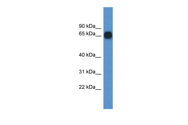
| Calculated MW | 62kDa |
| Gene ID | 12891 |
|---|---|
| Alias Symbol | AU067659, BB076446 |
| Other Names | Copine-6, Copine VI, Neuronal-copine, N-copine, Cpne6 |
| Format | Liquid. Purified antibody supplied in 1x PBS buffer with 0.09% (w/v) sodium azide and 2% sucrose. |
| Reconstitution & Storage | Add 50 ul of distilled water. Final anti-Cpne6 antibody concentration is 1 mg/ml in PBS buffer with 2% sucrose. For longer periods of storage, store at 20°C. Avoid repeat freeze-thaw cycles. |
| Precautions | Cpne6 antibody - C-terminal region is for research use only and not for use in diagnostic or therapeutic procedures. |
| Name | Cpne6 {ECO:0000312|MGI:MGI:1334445} |
|---|---|
| Function | Calcium-dependent phospholipid-binding protein that plays a role in calcium-mediated intracellular processes. Binds phospholipid membranes in a calcium-dependent manner (PubMed:9886090). Plays a role in dendrite formation by melanocytes (By similarity). |
| Cellular Location | Cytoplasm. Cell membrane. Endosome. Cytoplasmic vesicle, clathrin-coated vesicle. Perikaryon. Cell projection, dendrite. Note=Mainly cytoplasmic in absence of calcium. Associated predominantly with membranes in presence of calcium (PubMed:9886090). Translocates to the cell membrane in a calcium- dependent manner (PubMed:21087455, PubMed:26175110). Colocalized with transferrin in intracellular clathrin-coated membrane vesicles in a calcium-dependent manner (PubMed:21087455) |
| Tissue Location | Expressed in the brain (PubMed:9645480). Expressed in pyramidal cells, granule cells, and neurons in the dentate gyrus of the hippocampus and in granule cells of the olfactory bulb (at protein level). Expressed in pyramidal cells of the CA1-CA3 regions, in granule cells of the dentate gyrus, in granule cells of the olfactory bulbs, in the mitral cell layer and in neurons of the cerebral cortex layer II, brainstem and spinal cord (PubMed:9886090). Not detected in glial cells (PubMed:9645480, PubMed:9886090). |
Research Areas
Citations (0)

Thousands of laboratories across the world have published research that depended on the performance of antibodies from Abcepta to advance their research. Check out links to articles that cite our products in major peer-reviewed journals, organized by research category.
Submit your citation using an Abcepta antibody to
info@abcepta.com, and receive a free "I Love Antibodies" mug.
info@abcepta.com, and receive a free "I Love Antibodies" mug.

Application Protocols
Provided below are standard protocols that you may find useful for product applications.
References
Nakayama T.,et al.FEBS Lett. 428:80-84(1998).
Abcepta welcomes feedback from its customers.
If you have used an Abcepta product and would like to share how it has performed, please click on the "Submit Review" button and provide the requested information. Our staff will examine and post your review and contact you if needed.
If you have any additional inquiries please email technical services at tech@abcepta.com.
$ 389.00
Cat# AI14400
Ordering Information
United States
AlbaniaAustraliaAustriaBelgiumBosnia & HerzegovinaBrazilBulgariaCanadaCentral AmericaChinaCroatiaCyprusCzech RepublicDenmarkEstoniaFinlandFranceGermanyGreeceHong KongHungaryIcelandIndiaIndonesiaIrelandIsraelItalyJapanLatviaLithuaniaLuxembourgMacedoniaMalaysiaMaltaMexicoNetherlandsNew ZealandNorwayPakistanPolandPortugalRomaniaSerbiaSingaporeSlovakiaSloveniaSouth AfricaSouth KoreaSpainSwedenSwitzerlandTaiwanTurkeyUnited KingdomUnited StatesVietnamWorldwideOthers
USA Headquarters
(888) 735-7227 / (858) 622-0099 or (858) 875-1900
Other Products
Shipping Information
Domestic orders (in stock items)
Shipped out the same day. Orders placed after 1 PM (PST) will ship out the next business day.
International orders
Contact your local distributors
