


 Foundational characteristics of cancer include proliferation, angiogenesis, migration, evasion of apoptosis, and cellular immortality. Find key markers for these cellular processes and antibodies to detect them.
 The SUMOplot™ Analysis Program predicts and scores sumoylation sites in your protein. SUMOylation is a post-translational modification involved in various cellular processes, such as nuclear-cytosolic transport, transcriptional regulation, apoptosis, protein stability, response to stress, and progression through the cell cycle.
 The Autophagy Receptor Motif Plotter predicts and scores autophagy receptor binding sites in your protein. Identifying proteins connected to this pathway is critical to understanding the role of autophagy in physiological as well as pathological processes such as development, differentiation, neurodegenerative diseases, stress, infection, and cancer.
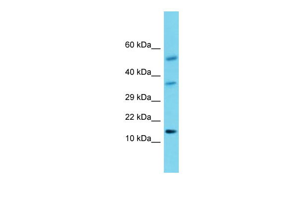
Hcrt Antibody - middle region
Rabbit Polyclonal Antibody
- SPECIFICATION
- CITATIONS
- PROTOCOLS
- BACKGROUND

Application
| WB |
|---|---|
| Primary Accession | O55232 |
| Other Accession | NM_013179, NP_037311 |
| Reactivity | Human, Mouse, Rat, Rabbit, Pig, Sheep, Horse, Bovine, Guinea Pig, Dog |
| Predicted | Human, Mouse, Rat, Rabbit, Pig, Sheep, Horse, Bovine, Guinea Pig, Dog |
| Host | Rabbit |
| Clonality | Polyclonal |
| Calculated MW | 14kDa |
| Gene ID | 25723 |
|---|---|
| Alias Symbol | orexin-A |
| Other Names | Orexin, Hypocretin, Hcrt, Orexin-A, Hypocretin-1, Hcrt1, Orexin-B, Hypocretin-2, Hcrt2, Hcrt, Ox, Ppox |
| Format | Liquid. Purified antibody supplied in 1x PBS buffer with 0.09% (w/v) sodium azide and 2% sucrose. |
| Reconstitution & Storage | Add 50 ul of distilled water. Final anti-Hcrt antibody concentration is 1 mg/ml in PBS buffer with 2% sucrose. For longer periods of storage, store at 20°C. Avoid repeat freeze-thaw cycles. |
| Precautions | Hcrt Antibody - middle region is for research use only and not for use in diagnostic or therapeutic procedures. |
| Name | Hcrt {ECO:0000312|RGD:2786} |
|---|---|
| Synonyms | Ox, Ppox |
| Function | Neuropeptides that play a significant role in the regulation of food intake and sleep-wakefulness, possibly by coordinating the complex behavioral and physiologic responses of these complementary homeostatic functions (PubMed:11283317, PubMed:11340621, PubMed:9491897). A broader role in the homeostatic regulation of energy metabolism, autonomic function, hormonal balance and the regulation of body fluids, is also suggested (PubMed:9491897). A modulation effect on luteinizing hormone-releasing hormone (LHRH) secretion also suggests a more minor contribution to the regulation of reproductive function (PubMed:11340621, PubMed:9879756). |
| Cellular Location | Rough endoplasmic reticulum. Cytoplasmic vesicle. Synapse Note=Associated with perikaryal rough endoplasmic reticulum as well as cytoplasmic large granular vesicles at synapses |
| Tissue Location | Mainly expressed in the brain, with expression in neurons in the dorsal lateral hypothalamic area (at protein level) (PubMed:9419374). Produced by a small group of neurons restricted to the lateral and posterior hypothalamus and perifornical areas (PubMed:9491897). Positive neurons project widely throughout the entire neuroaxis (PubMed:9491897). Particularly abundant projections in the cerebral cortex, olfactory bulb, hippocampus, amygdala, septum, diagonal band of Broca, bed nucleus of the stria terminalis, thalamus, anterior and posterior hypothalamus, midbrain, brainstem, and spinal cord (PubMed:9491897). Also expressed in the enteric nervous system and pancreas (PubMed:9491897). In small amount, also detected in the testis (PubMed:9491897). |

Thousands of laboratories across the world have published research that depended on the performance of antibodies from Abcepta to advance their research. Check out links to articles that cite our products in major peer-reviewed journals, organized by research category.
info@abcepta.com, and receive a free "I Love Antibodies" mug.

Provided below are standard protocols that you may find useful for product applications.
References
Sakurai T.,et al.Cell 92:573-585(1998).
de Lecea L.,et al.Proc. Natl. Acad. Sci. U.S.A. 95:322-327(1998).
Hungs M.,et al.Bioessays 23:397-408(2001).
Willie J.T.,et al.Annu. Rev. Neurosci. 24:429-458(2001).
If you have used an Abcepta product and would like to share how it has performed, please click on the "Submit Review" button and provide the requested information. Our staff will examine and post your review and contact you if needed.
If you have any additional inquiries please email technical services at tech@abcepta.com.
Ordering Information
Other Products
Shipping Information
