


 Foundational characteristics of cancer include proliferation, angiogenesis, migration, evasion of apoptosis, and cellular immortality. Find key markers for these cellular processes and antibodies to detect them.
 The SUMOplot™ Analysis Program predicts and scores sumoylation sites in your protein. SUMOylation is a post-translational modification involved in various cellular processes, such as nuclear-cytosolic transport, transcriptional regulation, apoptosis, protein stability, response to stress, and progression through the cell cycle.
 The Autophagy Receptor Motif Plotter predicts and scores autophagy receptor binding sites in your protein. Identifying proteins connected to this pathway is critical to understanding the role of autophagy in physiological as well as pathological processes such as development, differentiation, neurodegenerative diseases, stress, infection, and cancer.
ZNHIT2 Antibody - C-terminal region
Rabbit Polyclonal Antibody
- SPECIFICATION
- CITATIONS
- PROTOCOLS
- BACKGROUND

Application
| WB |
|---|---|
| Primary Accession | Q9UHR6 |
| Other Accession | NM_014205, NP_055020 |
| Reactivity | Human, Mouse, Rat, Rabbit, Pig, Yeast, Bovine, Guinea Pig, Dog |
| Predicted | Human, Rat, Rabbit, Pig, Bovine, Guinea Pig |
| Host | Rabbit |
| Clonality | Polyclonal |
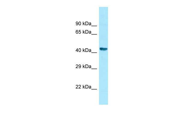
| Calculated MW | 43kDa |
| Gene ID | 741 |
|---|---|
| Alias Symbol | C11orf5, FON |
| Other Names | Zinc finger HIT domain-containing protein 2, Protein FON, ZNHIT2, C11orf5 |
| Format | Liquid. Purified antibody supplied in 1x PBS buffer with 0.09% (w/v) sodium azide and 2% sucrose. |
| Reconstitution & Storage | Add 50 ul of distilled water. Final anti-ZNHIT2 antibody concentration is 1 mg/ml in PBS buffer with 2% sucrose. For longer periods of storage, store at 20°C. Avoid repeat freeze-thaw cycles. |
| Precautions | ZNHIT2 Antibody - C-terminal region is for research use only and not for use in diagnostic or therapeutic procedures. |
| Name | ZNHIT2 |
|---|---|
| Synonyms | C11orf5 |
| Function | May act as a bridging factor mediating the interaction between the R2TP/Prefoldin-like (R2TP/PFDL) complex and U5 small nuclear ribonucleoprotein (U5 snRNP) (PubMed:28561026). Required for the interaction of R2TP complex subunit RPAP3 and prefoldin-like subunit URI1 with U5 snRNP proteins EFTUD2 and PRPF8 (PubMed:28561026). May play a role in regulating the composition of the U5 snRNP complex (PubMed:28561026). |
| Tissue Location | Low expression in most tissues; highly expressed in testis. |

Thousands of laboratories across the world have published research that depended on the performance of antibodies from Abcepta to advance their research. Check out links to articles that cite our products in major peer-reviewed journals, organized by research category.
info@abcepta.com, and receive a free "I Love Antibodies" mug.

Provided below are standard protocols that you may find useful for product applications.
References
Lemmens I.H.,et al.Mamm. Genome 11:78-80(2000).
Daub H.,et al.Mol. Cell 31:438-448(2008).
Dephoure N.,et al.Proc. Natl. Acad. Sci. U.S.A. 105:10762-10767(2008).
Burkard T.R.,et al.BMC Syst. Biol. 5:17-17(2011).
Van Damme P.,et al.Proc. Natl. Acad. Sci. U.S.A. 109:12449-12454(2012).
If you have used an Abcepta product and would like to share how it has performed, please click on the "Submit Review" button and provide the requested information. Our staff will examine and post your review and contact you if needed.
If you have any additional inquiries please email technical services at tech@abcepta.com.
Ordering Information
Other Products
Shipping Information
