


 Foundational characteristics of cancer include proliferation, angiogenesis, migration, evasion of apoptosis, and cellular immortality. Find key markers for these cellular processes and antibodies to detect them.
 The SUMOplot™ Analysis Program predicts and scores sumoylation sites in your protein. SUMOylation is a post-translational modification involved in various cellular processes, such as nuclear-cytosolic transport, transcriptional regulation, apoptosis, protein stability, response to stress, and progression through the cell cycle.
 The Autophagy Receptor Motif Plotter predicts and scores autophagy receptor binding sites in your protein. Identifying proteins connected to this pathway is critical to understanding the role of autophagy in physiological as well as pathological processes such as development, differentiation, neurodegenerative diseases, stress, infection, and cancer.
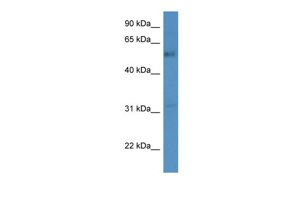
FAM126A antibody - C-terminal region
Rabbit Polyclonal Antibody
- SPECIFICATION
- CITATIONS
- PROTOCOLS
- BACKGROUND

Application
| WB |
|---|---|
| Primary Accession | Q9BYI3 |
| Other Accession | NM_032581, NP_115970 |
| Reactivity | Human, Mouse, Rat, Rabbit, Horse, Bovine, Guinea Pig, Dog |
| Predicted | Mouse, Rat, Bovine, Dog |
| Host | Rabbit |
| Clonality | Polyclonal |
| Calculated MW | 57kDa |
| Gene ID | 84668 |
|---|---|
| Alias Symbol | DRCTNNB1A, HCC, HLD5, HYCC1 |
| Other Names | Hyccin, Down-regulated by CTNNB1 protein A, Protein FAM126A, FAM126A, DRCTNNB1A |
| Format | Liquid. Purified antibody supplied in 1x PBS buffer with 0.09% (w/v) sodium azide and 2% sucrose. |
| Reconstitution & Storage | Add 50 ul of distilled water. Final anti-FAM126A antibody concentration is 1 mg/ml in PBS buffer with 2% sucrose. For longer periods of storage, store at 20°C. Avoid repeat freeze-thaw cycles. |
| Precautions | FAM126A antibody - C-terminal region is for research use only and not for use in diagnostic or therapeutic procedures. |
| Name | HYCC1 (HGNC:24587) |
|---|---|
| Function | Component of a complex required to localize phosphatidylinositol 4-kinase (PI4K) to the plasma membrane (PubMed:26571211). The complex acts as a regulator of phosphatidylinositol 4-phosphate (PtdIns(4)P) synthesis (PubMed:26571211). HYCC1 plays a key role in oligodendrocytes formation, a cell type with expanded plasma membrane that requires generation of PtdIns(4)P (PubMed:26571211). Its role in oligodendrocytes formation probably explains its importance in myelination of the central and peripheral nervous system (PubMed:16951682, PubMed:26571211). May also have a role in the beta- catenin/Lef signaling pathway (Probable). |
| Cellular Location | Cytoplasm, cytosol. Cell membrane Note=Localizes to the cytosol and is recruited to the plasma membrane following interaction with other components of the phosphatidylinositol 4-kinase (PI4K) complex. |
| Tissue Location | Widely expressed. Highest levels in heart, brain, placenta, spleen and testis. |

Thousands of laboratories across the world have published research that depended on the performance of antibodies from Abcepta to advance their research. Check out links to articles that cite our products in major peer-reviewed journals, organized by research category.
info@abcepta.com, and receive a free "I Love Antibodies" mug.

Provided below are standard protocols that you may find useful for product applications.
References
Kawasoe T.,et al.Cancer Res. 60:3354-3358(2000).
Ota T.,et al.Nat. Genet. 36:40-45(2004).
Bechtel S.,et al.BMC Genomics 8:399-399(2007).
Scherer S.W.,et al.Science 300:767-772(2003).
Mural R.J.,et al.Submitted (JUL-2005) to the EMBL/GenBank/DDBJ databases.
If you have used an Abcepta product and would like to share how it has performed, please click on the "Submit Review" button and provide the requested information. Our staff will examine and post your review and contact you if needed.
If you have any additional inquiries please email technical services at tech@abcepta.com.
Ordering Information
Shipping Information
