


 Foundational characteristics of cancer include proliferation, angiogenesis, migration, evasion of apoptosis, and cellular immortality. Find key markers for these cellular processes and antibodies to detect them.
 The SUMOplot™ Analysis Program predicts and scores sumoylation sites in your protein. SUMOylation is a post-translational modification involved in various cellular processes, such as nuclear-cytosolic transport, transcriptional regulation, apoptosis, protein stability, response to stress, and progression through the cell cycle.
 The Autophagy Receptor Motif Plotter predicts and scores autophagy receptor binding sites in your protein. Identifying proteins connected to this pathway is critical to understanding the role of autophagy in physiological as well as pathological processes such as development, differentiation, neurodegenerative diseases, stress, infection, and cancer.
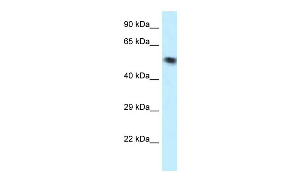
VPS4B antibody - N-terminal region
Rabbit Polyclonal Antibody
- SPECIFICATION
- CITATIONS
- PROTOCOLS
- BACKGROUND

Application
| WB |
|---|---|
| Primary Accession | O75351 |
| Other Accession | NM_004869, NP_004860 |
| Reactivity | Human, Rat, Rabbit, Pig, Horse, Bovine, Guinea Pig, Dog |
| Predicted | Human, Rabbit, Pig, Bovine, Guinea Pig, Dog |
| Host | Rabbit |
| Clonality | Polyclonal |
| Calculated MW | 49kDa |
| Gene ID | 9525 |
|---|---|
| Alias Symbol | MIG1, SKD1, SKD1B, VPS4-2 |
| Other Names | Vacuolar protein sorting-associated protein 4B, 3.6.4.6, Cell migration-inducing gene 1 protein, Suppressor of K(+) transport growth defect 1, Protein SKD1, VPS4B, SKD1, VPS42 |
| Format | Liquid. Purified antibody supplied in 1x PBS buffer with 0.09% (w/v) sodium azide and 2% sucrose. |
| Reconstitution & Storage | Add 50 ul of distilled water. Final anti-VPS4B antibody concentration is 1 mg/ml in PBS buffer with 2% sucrose. For longer periods of storage, store at 20°C. Avoid repeat freeze-thaw cycles. |
| Precautions | VPS4B antibody - N-terminal region is for research use only and not for use in diagnostic or therapeutic procedures. |
| Name | VPS4B (HGNC:10895) |
|---|---|
| Synonyms | SKD1, VPS42 |
| Function | Involved in late steps of the endosomal multivesicular bodies (MVB) pathway. Recognizes membrane-associated ESCRT-III assemblies and catalyzes their ATP-dependent disassembly, possibly in combination with membrane fission (PubMed:18687924). Redistributes the ESCRT-III components to the cytoplasm for further rounds of MVB sorting. MVBs contain intraluminal vesicles (ILVs) that are generated by invagination and scission from the limiting membrane of the endosome and mostly are delivered to lysosomes enabling degradation of membrane proteins, such as stimulated growth factor receptors, lysosomal enzymes and lipids. VPS4A/B are required for the exosomal release of SDCBP, CD63 and syndecan (PubMed:22660413). |
| Cellular Location | Late endosome membrane {ECO:0000250|UniProtKB:P46467}; Peripheral membrane protein. Note=Membrane-associated in the prevacuolar endosomal compartment. Localized in HIV-1 particles purified from acutely infected cells. |
| Tissue Location | Ubiquitously expressed. |

Thousands of laboratories across the world have published research that depended on the performance of antibodies from Abcepta to advance their research. Check out links to articles that cite our products in major peer-reviewed journals, organized by research category.
info@abcepta.com, and receive a free "I Love Antibodies" mug.

Provided below are standard protocols that you may find useful for product applications.
References
Scheuring S.,et al.J. Mol. Biol. 312:469-480(2001).
Beyer A.,et al.Gene 305:47-59(2003).
Mao M.,et al.Proc. Natl. Acad. Sci. U.S.A. 95:8175-8180(1998).
Kim J.W.,et al.Submitted (FEB-2003) to the EMBL/GenBank/DDBJ databases.
Mural R.J.,et al.Submitted (JUL-2005) to the EMBL/GenBank/DDBJ databases.
If you have used an Abcepta product and would like to share how it has performed, please click on the "Submit Review" button and provide the requested information. Our staff will examine and post your review and contact you if needed.
If you have any additional inquiries please email technical services at tech@abcepta.com.
Ordering Information
Other Products
Shipping Information
