


 Foundational characteristics of cancer include proliferation, angiogenesis, migration, evasion of apoptosis, and cellular immortality. Find key markers for these cellular processes and antibodies to detect them.
 The SUMOplot™ Analysis Program predicts and scores sumoylation sites in your protein. SUMOylation is a post-translational modification involved in various cellular processes, such as nuclear-cytosolic transport, transcriptional regulation, apoptosis, protein stability, response to stress, and progression through the cell cycle.
 The Autophagy Receptor Motif Plotter predicts and scores autophagy receptor binding sites in your protein. Identifying proteins connected to this pathway is critical to understanding the role of autophagy in physiological as well as pathological processes such as development, differentiation, neurodegenerative diseases, stress, infection, and cancer.
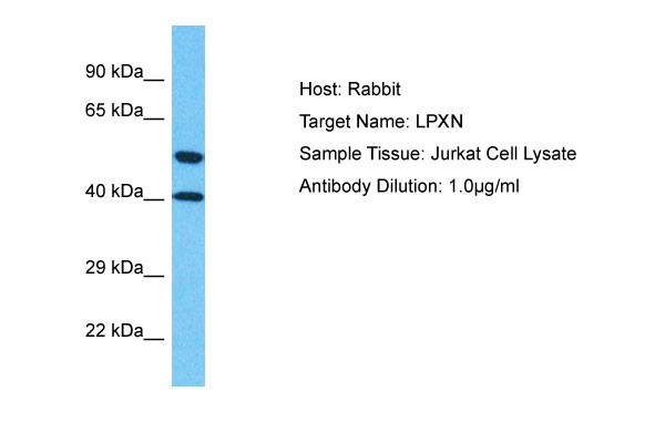
LPXN Antibody - C-terminal region
Rabbit Polyclonal Antibody
- SPECIFICATION
- CITATIONS
- PROTOCOLS
- BACKGROUND

Application
| WB |
|---|---|
| Primary Accession | O60711 |
| Other Accession | NP_004802 |
| Reactivity | Human |
| Host | Rabbit |
| Clonality | Polyclonal |
| Calculated MW | 42kDa |
| Gene ID | 9404 |
|---|---|
| Alias Symbol | LPXN, LDLP, |
| Other Names | Leupaxin, LPXN, LDLP |
| Format | Liquid. Purified antibody supplied in 1x PBS buffer with 0.09% (w/v) sodium azide and 2% sucrose. |
| Reconstitution & Storage | Add 50 &mu, l of distilled water. Final Anti-LPXN antibody concentration is 1 mg/ml in PBS buffer with 2% sucrose. For longer periods of storage, store at -20°C. Avoid repeat freeze-thaw cycles. |
| Precautions | LPXN Antibody - C-terminal region is for research use only and not for use in diagnostic or therapeutic procedures. |
| Name | LPXN |
|---|---|
| Synonyms | LDLP |
| Function | Transcriptional coactivator for androgen receptor (AR) and serum response factor (SRF). Contributes to the regulation of cell adhesion, spreading and cell migration and acts as a negative regulator in integrin-mediated cell adhesion events. Suppresses the integrin- induced tyrosine phosphorylation of paxillin (PXN). May play a critical role as an adapter protein in the formation of the adhesion zone in osteoclasts. Negatively regulates B-cell antigen receptor (BCR) signaling. |
| Cellular Location | Cytoplasm. Cell junction, focal adhesion. Nucleus. Cytoplasm, perinuclear region. Cell projection, podosome. Cell membrane. Note=Shuttles between the cytoplasm and nucleus. Recruited to the cell membrane following B-cell antigen receptor (BCR) cross-linking in B-cells. Enhanced focal adhesion kinase activity (PTK2/FAK) attenuates its nuclear accumulation and limits its ability to enhance serum response factor (SRF)-dependent gene transcription. Targeting to focal adhesions is essential for its tyrosine phosphorylation in response to bombesin |
| Tissue Location | Macrophages, monocytes and osteoclasts (at protein level). Strongly expressed in cells and tissues of hematopoietic origin. Highest expression in lymphoid tissues such as spleen, lymph node, thymus and appendix and in the vascular smooth muscle. Lower levels in bone marrow and fetal liver. Also expressed in peripheral blood lymphocytes and a number of hematopoietic cell lines. Very low levels found in epithelial cell lines. Expressed in prostate cancer (PCa) cells and its expression intensity is directly linked to PCa progression. |

Thousands of laboratories across the world have published research that depended on the performance of antibodies from Abcepta to advance their research. Check out links to articles that cite our products in major peer-reviewed journals, organized by research category.
info@abcepta.com, and receive a free "I Love Antibodies" mug.

Provided below are standard protocols that you may find useful for product applications.
References
Lipsky B.P.,et al.J. Biol. Chem. 273:11709-11713(1998).
Ota T.,et al.Nat. Genet. 36:40-45(2004).
Ebert L.,et al.Submitted (JUN-2004) to the EMBL/GenBank/DDBJ databases.
Suzuki Y.,et al.Submitted (APR-2005) to the EMBL/GenBank/DDBJ databases.
Taylor T.D.,et al.Nature 440:497-500(2006).
If you have used an Abcepta product and would like to share how it has performed, please click on the "Submit Review" button and provide the requested information. Our staff will examine and post your review and contact you if needed.
If you have any additional inquiries please email technical services at tech@abcepta.com.
Ordering Information
Other Products
Shipping Information
