


 Foundational characteristics of cancer include proliferation, angiogenesis, migration, evasion of apoptosis, and cellular immortality. Find key markers for these cellular processes and antibodies to detect them.
 The SUMOplot™ Analysis Program predicts and scores sumoylation sites in your protein. SUMOylation is a post-translational modification involved in various cellular processes, such as nuclear-cytosolic transport, transcriptional regulation, apoptosis, protein stability, response to stress, and progression through the cell cycle.
 The Autophagy Receptor Motif Plotter predicts and scores autophagy receptor binding sites in your protein. Identifying proteins connected to this pathway is critical to understanding the role of autophagy in physiological as well as pathological processes such as development, differentiation, neurodegenerative diseases, stress, infection, and cancer.
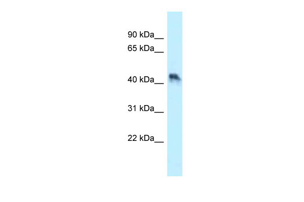
ENTPD5 antibody - N-terminal region
Rabbit Polyclonal Antibody
- SPECIFICATION
- CITATIONS
- PROTOCOLS
- BACKGROUND

Application
| WB |
|---|---|
| Primary Accession | O75356 |
| Other Accession | NM_001249, NP_001240 |
| Reactivity | Human, Mouse, Rat, Rabbit, Pig, Horse, Bovine, Guinea Pig, Dog |
| Predicted | Human, Mouse, Rat, Rabbit, Pig, Horse, Bovine, Guinea Pig, Dog |
| Host | Rabbit |
| Clonality | Polyclonal |
| Calculated MW | 47kDa |
| Gene ID | 957 |
|---|---|
| Alias Symbol | CD39L4, MGC163357, MGC163359, NTPDase-5, PCPH |
| Other Names | Ectonucleoside triphosphate diphosphohydrolase 5, NTPDase 5, 3.6.1.6, CD39 antigen-like 4, ER-UDPase, Guanosine-diphosphatase ENTPD5, GDPase ENTPD5, 3.6.1.42, Nucleoside diphosphatase, Uridine-diphosphatase ENTPD5, UDPase ENTPD5, ENTPD5, CD39L4, PCPH |
| Format | Liquid. Purified antibody supplied in 1x PBS buffer with 0.09% (w/v) sodium azide and 2% sucrose. |
| Reconstitution & Storage | Add 50 ul of distilled water. Final anti-ENTPD5 antibody concentration is 1 mg/ml in PBS buffer with 2% sucrose. For longer periods of storage, store at 20°C. Avoid repeat freeze-thaw cycles. |
| Precautions | ENTPD5 antibody - N-terminal region is for research use only and not for use in diagnostic or therapeutic procedures. |
| Name | ENTPD5 (HGNC:3367) |
|---|---|
| Function | Hydrolyzes nucleoside diphosphates with a preference for GDP, IDP and UDP compared to ADP and CDP (PubMed:10400613, PubMed:15698960). In the lumen of the endoplasmic reticulum, hydrolyzes UDP that acts as an end-product feedback inhibitor of the UDP-Glc:glycoprotein glucosyltransferases. UMP can be transported back by an UDP-sugar antiporter to the cytosol where it is consumed to regenerate UDP- glucose. Therefore, it positively regulates protein reglucosylation by clearing UDP from the ER lumen and by promoting the regeneration of UDP-glucose. Protein reglucosylation is essential to proper glycoprotein folding and quality control in the ER (By similarity). |
| Cellular Location | Endoplasmic reticulum {ECO:0000250|UniProtKB:Q9WUZ9}. Secreted |
| Tissue Location | Expressed in adult liver, kidney, prostate, testis and colon. Much weaker expression in other tissues |

Thousands of laboratories across the world have published research that depended on the performance of antibodies from Abcepta to advance their research. Check out links to articles that cite our products in major peer-reviewed journals, organized by research category.
info@abcepta.com, and receive a free "I Love Antibodies" mug.

Provided below are standard protocols that you may find useful for product applications.
References
Chadwick B.P.,et al.Genomics 50:357-367(1998).
Recio J.A.,et al.Mol. Carcinog. 27:229-236(2000).
Murphy-Piedmonte D.M.,et al.Biochim. Biophys. Acta 1747:251-259(2005).
Heilig R.,et al.Nature 421:601-607(2003).
Mural R.J.,et al.Submitted (JUL-2005) to the EMBL/GenBank/DDBJ databases.
If you have used an Abcepta product and would like to share how it has performed, please click on the "Submit Review" button and provide the requested information. Our staff will examine and post your review and contact you if needed.
If you have any additional inquiries please email technical services at tech@abcepta.com.
Ordering Information
Other Products
Shipping Information
