


 Foundational characteristics of cancer include proliferation, angiogenesis, migration, evasion of apoptosis, and cellular immortality. Find key markers for these cellular processes and antibodies to detect them.
 The SUMOplot™ Analysis Program predicts and scores sumoylation sites in your protein. SUMOylation is a post-translational modification involved in various cellular processes, such as nuclear-cytosolic transport, transcriptional regulation, apoptosis, protein stability, response to stress, and progression through the cell cycle.
 The Autophagy Receptor Motif Plotter predicts and scores autophagy receptor binding sites in your protein. Identifying proteins connected to this pathway is critical to understanding the role of autophagy in physiological as well as pathological processes such as development, differentiation, neurodegenerative diseases, stress, infection, and cancer.
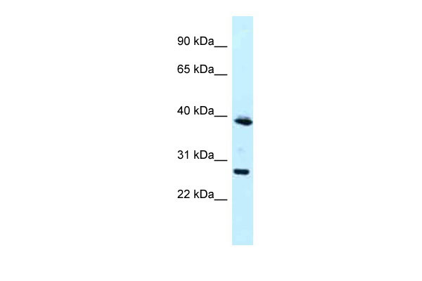
MAGEF1 antibody - N-terminal region
Rabbit Polyclonal Antibody
- SPECIFICATION
- CITATIONS
- PROTOCOLS
- BACKGROUND

Application
| WB |
|---|---|
| Primary Accession | Q9HAY2 |
| Other Accession | NM_022149, NP_071432 |
| Reactivity | Human |
| Predicted | Human |
| Host | Rabbit |
| Clonality | Polyclonal |
| Calculated MW | 34kDa |
| Gene ID | 64110 |
|---|---|
| Alias Symbol | MGC19617 |
| Other Names | Melanoma-associated antigen F1, MAGE-F1 antigen, MAGEF1 |
| Format | Liquid. Purified antibody supplied in 1x PBS buffer with 0.09% (w/v) sodium azide and 2% sucrose. |
| Reconstitution & Storage | Add 50 ul of distilled water. Final anti-MAGEF1 antibody concentration is 1 mg/ml in PBS buffer with 2% sucrose. For longer periods of storage, store at 20°C. Avoid repeat freeze-thaw cycles. |
| Precautions | MAGEF1 antibody - N-terminal region is for research use only and not for use in diagnostic or therapeutic procedures. |
| Name | MAGEF1 (HGNC:29639) |
|---|---|
| Function | Enhances ubiquitin ligase activity of RING-type zinc finger- containing E3 ubiquitin ligases. Proposed to act through recruitment and/or stabilization of the E2 ubiquitin-conjugating enzyme at the E3:substrate complex. MAGEF1-NSMCE1 ubiquitin ligase complex promotes proteasomal degradation of MMS19, a key component of the cytosolic iron-sulfur protein assembly (CIA) machinery. Down-regulation of MMS19 impairs the activity of several DNA repair and metabolism enzymes such as ERCC2/XPD, FANCJ, RTEL1 and POLD1 that require iron-sulfur clusters as cofactors. May negatively regulate genome integrity by inhibiting homologous recombination-mediated double-strand break DNA repair (PubMed:29225034). |
| Tissue Location | Ubiquitous.. |

Thousands of laboratories across the world have published research that depended on the performance of antibodies from Abcepta to advance their research. Check out links to articles that cite our products in major peer-reviewed journals, organized by research category.
info@abcepta.com, and receive a free "I Love Antibodies" mug.

Provided below are standard protocols that you may find useful for product applications.
References
Stone B.,et al.Gene 267:173-182(2001).
Muzny D.M.,et al.Nature 440:1194-1198(2006).
Chomez P.,et al.Cancer Res. 61:5544-5551(2001).
Doyle J.M.,et al.Mol. Cell 39:963-974(2010).
If you have used an Abcepta product and would like to share how it has performed, please click on the "Submit Review" button and provide the requested information. Our staff will examine and post your review and contact you if needed.
If you have any additional inquiries please email technical services at tech@abcepta.com.
Ordering Information
Other Products
Shipping Information
