


 Foundational characteristics of cancer include proliferation, angiogenesis, migration, evasion of apoptosis, and cellular immortality. Find key markers for these cellular processes and antibodies to detect them.
 The SUMOplot™ Analysis Program predicts and scores sumoylation sites in your protein. SUMOylation is a post-translational modification involved in various cellular processes, such as nuclear-cytosolic transport, transcriptional regulation, apoptosis, protein stability, response to stress, and progression through the cell cycle.
 The Autophagy Receptor Motif Plotter predicts and scores autophagy receptor binding sites in your protein. Identifying proteins connected to this pathway is critical to understanding the role of autophagy in physiological as well as pathological processes such as development, differentiation, neurodegenerative diseases, stress, infection, and cancer.
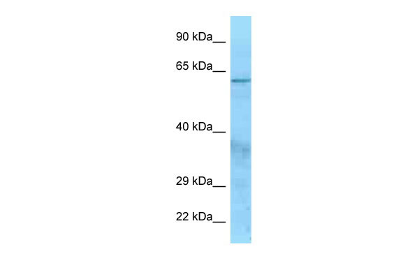
CFI antibody - N-terminal region
Rabbit Polyclonal Antibody
- SPECIFICATION
- CITATIONS
- PROTOCOLS
- BACKGROUND

Application
| WB |
|---|---|
| Primary Accession | P05156 |
| Other Accession | NM_000204, NP_000195 |
| Reactivity | Human |
| Predicted | Human |
| Host | Rabbit |
| Clonality | Polyclonal |
| Calculated MW | 64kDa |
| Gene ID | 3426 |
|---|---|
| Alias Symbol | AHUS3, C3BINA, C3b-INA, FI, IF, KAF |
| Other Names | Complement factor I, 3.4.21.45, C3B/C4B inactivator, Complement factor I heavy chain, Complement factor I light chain, CFI, IF |
| Format | Liquid. Purified antibody supplied in 1x PBS buffer with 0.09% (w/v) sodium azide and 2% sucrose. |
| Reconstitution & Storage | Add 50 ul of distilled water. Final anti-CFI antibody concentration is 1 mg/ml in PBS buffer with 2% sucrose. For longer periods of storage, store at 20°C. Avoid repeat freeze-thaw cycles. |
| Precautions | CFI antibody - N-terminal region is for research use only and not for use in diagnostic or therapeutic procedures. |
| Name | CFI |
|---|---|
| Synonyms | IF |
| Function | Trypsin-like serine protease that plays an essential role in regulating the immune response by controlling all complement pathways. Inhibits these pathways by cleaving three peptide bonds in the alpha- chain of C3b and two bonds in the alpha-chain of C4b thereby inactivating these proteins (PubMed:17320177, PubMed:7360115). Essential cofactors for these reactions include factor H and C4BP in the fluid phase and membrane cofactor protein/CD46 and CR1 on cell surfaces (PubMed:12055245, PubMed:2141838, PubMed:9605165). The presence of these cofactors on healthy cells allows degradation of deposited C3b by CFI in order to prevent undesired complement activation, while in apoptotic cells or microbes, the absence of such cofactors leads to C3b-mediated complement activation and subsequent opsonization (PubMed:28671664). |
| Cellular Location | Secreted, extracellular space. Secreted |
| Tissue Location | Expressed in the liver by hepatocytes (PubMed:6327681). Also present in other cells such as monocytes, fibroblasts or keratinocytes (PubMed:17320177, PubMed:6444659) |

Thousands of laboratories across the world have published research that depended on the performance of antibodies from Abcepta to advance their research. Check out links to articles that cite our products in major peer-reviewed journals, organized by research category.
info@abcepta.com, and receive a free "I Love Antibodies" mug.

Provided below are standard protocols that you may find useful for product applications.
References
Catterall C.F.,et al.Biochem. J. 242:849-856(1987).
Goldberger G.,et al.J. Biol. Chem. 262:10065-10071(1987).
Hillier L.W.,et al.Nature 434:724-731(2005).
Minta J.O.,et al.Gene 208:17-24(1998).
Ullman C.G.,et al.FEBS Lett. 371:199-203(1995).
If you have used an Abcepta product and would like to share how it has performed, please click on the "Submit Review" button and provide the requested information. Our staff will examine and post your review and contact you if needed.
If you have any additional inquiries please email technical services at tech@abcepta.com.
Ordering Information
Other Products
Shipping Information
