


 Foundational characteristics of cancer include proliferation, angiogenesis, migration, evasion of apoptosis, and cellular immortality. Find key markers for these cellular processes and antibodies to detect them.
 The SUMOplot™ Analysis Program predicts and scores sumoylation sites in your protein. SUMOylation is a post-translational modification involved in various cellular processes, such as nuclear-cytosolic transport, transcriptional regulation, apoptosis, protein stability, response to stress, and progression through the cell cycle.
 The Autophagy Receptor Motif Plotter predicts and scores autophagy receptor binding sites in your protein. Identifying proteins connected to this pathway is critical to understanding the role of autophagy in physiological as well as pathological processes such as development, differentiation, neurodegenerative diseases, stress, infection, and cancer.
Pdia3 antibody - N-terminal region
Rabbit Polyclonal Antibody
- SPECIFICATION
- CITATIONS
- PROTOCOLS
- BACKGROUND

Application
| WB |
|---|---|
| Primary Accession | P27773 |
| Other Accession | NM_007952, NP_031978 |
| Reactivity | Human, Mouse, Rat, Rabbit, Pig, Goat, Sheep, Horse, Bovine, Guinea Pig, Dog |
| Predicted | Human, Mouse, Rat, Rabbit, Pig, Chicken, Goat, Sheep, Bovine, Dog |
| Host | Rabbit |
| Clonality | Polyclonal |
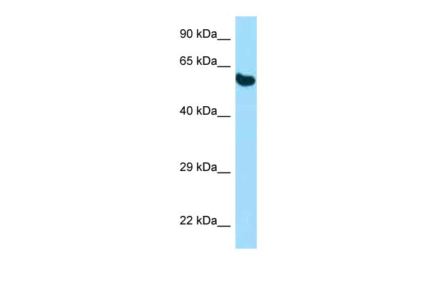
| Calculated MW | 57kDa |
| Gene ID | 14827 |
|---|---|
| Alias Symbol | 58kDa, ERp57, ERp60, ERp61, Erp, Grp58, PDI, PDI-Q2, PI-PLC, PLC[a], Plca |
| Other Names | Protein disulfide-isomerase A3, 5.3.4.1, 58 kDa glucose-regulated protein, 58 kDa microsomal protein, p58, Disulfide isomerase ER-60, Endoplasmic reticulum resident protein 57, ER protein 57, ERp57, Endoplasmic reticulum resident protein 60, ER protein 60, ERp60, Pdia3, Erp, Erp60, Grp58 |
| Format | Liquid. Purified antibody supplied in 1x PBS buffer with 0.09% (w/v) sodium azide and 2% sucrose. |
| Reconstitution & Storage | Add 50 ul of distilled water. Final anti-Pdia3 antibody concentration is 1 mg/ml in PBS buffer with 2% sucrose. For longer periods of storage, store at 20°C. Avoid repeat freeze-thaw cycles. |
| Precautions | Pdia3 antibody - N-terminal region is for research use only and not for use in diagnostic or therapeutic procedures. |
| Name | Pdia3 |
|---|---|
| Synonyms | Erp, Erp60, Grp58 |
| Function | Protein disulfide isomerase that catalyzes the formation, isomerization, and reduction or oxidation of disulfide bonds in client proteins and functions as a protein folding chaperone. Core component of the major histocompatibility complex class I (MHC I) peptide loading complex where it functions as an essential folding chaperone for TAPBP. Through TAPBP, assists the dynamic assembly of the MHC I complex with high affinity antigens in the endoplasmic reticulum. Therefore, plays a crucial role in the presentation of antigens to cytotoxic T cells in adaptive immunity. |
| Cellular Location | Endoplasmic reticulum {ECO:0000250|UniProtKB:P30101}. Endoplasmic reticulum lumen {ECO:0000250|UniProtKB:P11598}. Melanosome {ECO:0000250|UniProtKB:P30101} |
| Tissue Location | In caput and cauda epididymal spermatozoa, detected in the acrosome and principal piece (at protein level) |

Thousands of laboratories across the world have published research that depended on the performance of antibodies from Abcepta to advance their research. Check out links to articles that cite our products in major peer-reviewed journals, organized by research category.
info@abcepta.com, and receive a free "I Love Antibodies" mug.

Provided below are standard protocols that you may find useful for product applications.
References
Hempel W.M.,et al.J. Immunol. 146:3713-3720(1991).
Wang W.,et al.Submitted (APR-2005) to the EMBL/GenBank/DDBJ databases.
Carninci P.,et al.Science 309:1559-1563(2005).
Church D.M.,et al.PLoS Biol. 7:E1000112-E1000112(2009).
Merrick B.A.,et al.Electrophoresis 15:735-745(1994).
If you have used an Abcepta product and would like to share how it has performed, please click on the "Submit Review" button and provide the requested information. Our staff will examine and post your review and contact you if needed.
If you have any additional inquiries please email technical services at tech@abcepta.com.
Ordering Information
Other Products
Shipping Information
