


 Foundational characteristics of cancer include proliferation, angiogenesis, migration, evasion of apoptosis, and cellular immortality. Find key markers for these cellular processes and antibodies to detect them.
 The SUMOplot™ Analysis Program predicts and scores sumoylation sites in your protein. SUMOylation is a post-translational modification involved in various cellular processes, such as nuclear-cytosolic transport, transcriptional regulation, apoptosis, protein stability, response to stress, and progression through the cell cycle.
 The Autophagy Receptor Motif Plotter predicts and scores autophagy receptor binding sites in your protein. Identifying proteins connected to this pathway is critical to understanding the role of autophagy in physiological as well as pathological processes such as development, differentiation, neurodegenerative diseases, stress, infection, and cancer.
LILRB2 antibody - C-terminal region
Rabbit Polyclonal Antibody
- SPECIFICATION
- CITATIONS
- PROTOCOLS
- BACKGROUND

Application
| WB |
|---|---|
| Primary Accession | Q8N423 |
| Other Accession | NM_005874, NP_005865 |
| Reactivity | Human |
| Predicted | Human |
| Host | Rabbit |
| Clonality | Polyclonal |
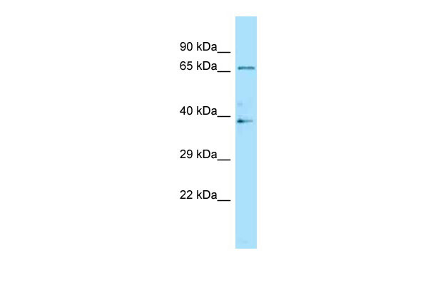
| Calculated MW | 65kDa |
| Gene ID | 10288 |
|---|---|
| Alias Symbol | CD85D, ILT4, LILRA6, LIR-2, LIR2, MIR-10, MIR10 |
| Other Names | Leukocyte immunoglobulin-like receptor subfamily B member 2, LIR-2, Leukocyte immunoglobulin-like receptor 2, CD85 antigen-like family member D, Immunoglobulin-like transcript 4, ILT-4, Monocyte/macrophage immunoglobulin-like receptor 10, MIR-10, CD85d, LILRB2, ILT4, LIR2, MIR10 |
| Format | Liquid. Purified antibody supplied in 1x PBS buffer with 0.09% (w/v) sodium azide and 2% sucrose. |
| Reconstitution & Storage | Add 50 ul of distilled water. Final anti-LILRB2 antibody concentration is 1 mg/ml in PBS buffer with 2% sucrose. For longer periods of storage, store at 20°C. Avoid repeat freeze-thaw cycles. |
| Precautions | LILRB2 antibody - C-terminal region is for research use only and not for use in diagnostic or therapeutic procedures. |
| Name | LILRB2 (HGNC:6606) |
|---|---|
| Function | Receptor for class I MHC antigens. Recognizes a broad spectrum of HLA-A, HLA-B, HLA-C, HLA-G and HLA-F alleles (PubMed:11169396, PubMed:12853576, PubMed:16455647, PubMed:20448110, PubMed:27859042). Involved in the down-regulation of the immune response and the development of tolerance. Recognizes HLA-G in complex with B2M/beta-2 microglobulin and a nonamer self-peptide (peptide-bound HLA-G-B2M) triggering differentiation of type 1 regulatory T cells and myeloid-derived suppressor cells, both of which actively maintain maternal-fetal tolerance (PubMed:16455647, PubMed:20448110, PubMed:27859042). Competes with CD8A for binding to class I MHC antigens. Inhibits FCGR1A-mediated phosphorylation of cellular proteins and mobilization of intracellular calcium ions (PubMed:11875462, PubMed:12853576, PubMed:9548455, PubMed:9842885). |
| Cellular Location | Cell membrane; Single-pass type I membrane protein |
| Tissue Location | Expressed in monocytes and at lower levels in myeloid and plasmacytoid dendritic cells. Expressed in tolerogenic IL10-producing dendritic cells (PubMed:20448110). Expressed in myeloid- derived suppressor cells during pregnancy (PubMed:27859042). Detected at low levels in natural killer (NK) cells. Expressed in B cells |

Thousands of laboratories across the world have published research that depended on the performance of antibodies from Abcepta to advance their research. Check out links to articles that cite our products in major peer-reviewed journals, organized by research category.
info@abcepta.com, and receive a free "I Love Antibodies" mug.

Provided below are standard protocols that you may find useful for product applications.
References
Colonna M.,et al.J. Exp. Med. 186:1809-1818(1997).
Borges L.,et al.J. Immunol. 159:5192-5196(1997).
Canavez F.C.,et al.Submitted (JUL-2000) to the EMBL/GenBank/DDBJ databases.
Grimwood J.,et al.Nature 428:529-535(2004).
Fanger N.A.,et al.Eur. J. Immunol. 28:3423-3434(1998).
If you have used an Abcepta product and would like to share how it has performed, please click on the "Submit Review" button and provide the requested information. Our staff will examine and post your review and contact you if needed.
If you have any additional inquiries please email technical services at tech@abcepta.com.
Ordering Information
Other Products
Shipping Information
