


 Foundational characteristics of cancer include proliferation, angiogenesis, migration, evasion of apoptosis, and cellular immortality. Find key markers for these cellular processes and antibodies to detect them.
 The SUMOplot™ Analysis Program predicts and scores sumoylation sites in your protein. SUMOylation is a post-translational modification involved in various cellular processes, such as nuclear-cytosolic transport, transcriptional regulation, apoptosis, protein stability, response to stress, and progression through the cell cycle.
 The Autophagy Receptor Motif Plotter predicts and scores autophagy receptor binding sites in your protein. Identifying proteins connected to this pathway is critical to understanding the role of autophagy in physiological as well as pathological processes such as development, differentiation, neurodegenerative diseases, stress, infection, and cancer.
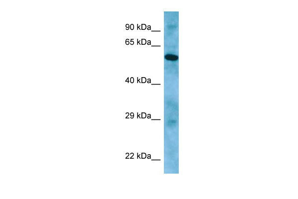
APLF Antibody - C-terminal region
Rabbit Polyclonal Antibody
- SPECIFICATION
- CITATIONS
- PROTOCOLS
- BACKGROUND

Application
| WB |
|---|---|
| Primary Accession | Q8IW19 |
| Other Accession | NM_173545, NP_775816 |
| Reactivity | Human, Mouse, Rat, Rabbit, Pig, Horse, Bovine |
| Predicted | Human, Mouse, Rat, Rabbit, Pig, Horse, Bovine |
| Host | Rabbit |
| Clonality | Polyclonal |
| Calculated MW | 57kDa |
| Gene ID | 200558 |
|---|---|
| Alias Symbol | APFL, C2orf13, FLJ16593, MGC47799, PALF, Xip1 |
| Other Names | Aprataxin and PNK-like factor, 4.2.99.18, Apurinic-apyrimidinic endonuclease APLF, PNK and APTX-like FHA domain-containing protein, XRCC1-interacting protein 1, APLF, C2orf13, PALF, XIP1 |
| Format | Liquid. Purified antibody supplied in 1x PBS buffer with 0.09% (w/v) sodium azide and 2% sucrose. |
| Reconstitution & Storage | Add 50 ul of distilled water. Final anti-APLF antibody concentration is 1 mg/ml in PBS buffer with 2% sucrose. For longer periods of storage, store at 20°C. Avoid repeat freeze-thaw cycles. |
| Precautions | APLF Antibody - C-terminal region is for research use only and not for use in diagnostic or therapeutic procedures. |
| Name | APLF {ECO:0000303|PubMed:17353262, ECO:0000312|HGNC:HGNC:28724} |
|---|---|
| Function | Histone chaperone involved in single-strand and double-strand DNA break repair (PubMed:17353262, PubMed:17396150, PubMed:21211721, PubMed:21211722, PubMed:29905837, PubMed:30104678). Recruited to sites of DNA damage through interaction with branched poly-ADP-ribose chains, a polymeric post-translational modification synthesized transiently at sites of chromosomal damage to accelerate DNA strand break repair reactions (PubMed:17353262, PubMed:17396150, PubMed:21211721, PubMed:30104678). Following recruitment to DNA damage sites, acts as a histone chaperone that mediates histone eviction during DNA repair and promotes recruitment of histone variant MACROH2A1 (PubMed:21211722, PubMed:29905837, PubMed:30104678). Also has a nuclease activity: displays apurinic-apyrimidinic (AP) endonuclease and 3'-5' exonuclease activities in vitro (PubMed:17353262, PubMed:17396150). Also able to introduce nicks at hydroxyuracil and other types of pyrimidine base damage (PubMed:17353262, PubMed:17396150). Together with PARP3, promotes the retention of the LIG4-XRCC4 complex on chromatin and accelerate DNA ligation during non-homologous end-joining (NHEJ) (PubMed:21211721, PubMed:23689425). Also acts as a negative regulator of cell pluripotency by promoting histone exchange (By similarity). Required for the embryo implantation during the epithelial to mesenchymal transition in females (By similarity). |
| Cellular Location | Nucleus. Chromosome. Cytoplasm, cytosol. Note=Localizes to DNA damage sites (PubMed:18172500, PubMed:18474613, PubMed:21211721, PubMed:21211722, PubMed:23689425). Accumulates at single-strand breaks and double-strand breaks via the PBZ-type zinc fingers (PubMed:18172500) |

Thousands of laboratories across the world have published research that depended on the performance of antibodies from Abcepta to advance their research. Check out links to articles that cite our products in major peer-reviewed journals, organized by research category.
info@abcepta.com, and receive a free "I Love Antibodies" mug.

Provided below are standard protocols that you may find useful for product applications.
References
Ota T.,et al.Nat. Genet. 36:40-45(2004).
Hillier L.W.,et al.Nature 434:724-731(2005).
Kanno S.,et al.EMBO J. 26:2094-2103(2007).
Bekker-Jensen S.,et al.J. Biol. Chem. 282:19638-19643(2007).
Iles N.,et al.Mol. Cell. Biol. 27:3793-3803(2007).
If you have used an Abcepta product and would like to share how it has performed, please click on the "Submit Review" button and provide the requested information. Our staff will examine and post your review and contact you if needed.
If you have any additional inquiries please email technical services at tech@abcepta.com.
Ordering Information
Other Products
Shipping Information
