


 Foundational characteristics of cancer include proliferation, angiogenesis, migration, evasion of apoptosis, and cellular immortality. Find key markers for these cellular processes and antibodies to detect them.
 The SUMOplot™ Analysis Program predicts and scores sumoylation sites in your protein. SUMOylation is a post-translational modification involved in various cellular processes, such as nuclear-cytosolic transport, transcriptional regulation, apoptosis, protein stability, response to stress, and progression through the cell cycle.
 The Autophagy Receptor Motif Plotter predicts and scores autophagy receptor binding sites in your protein. Identifying proteins connected to this pathway is critical to understanding the role of autophagy in physiological as well as pathological processes such as development, differentiation, neurodegenerative diseases, stress, infection, and cancer.
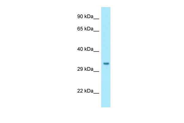
NIPSNAP1 Antibody - C-terminal region
Rabbit Polyclonal Antibody
- SPECIFICATION
- CITATIONS
- PROTOCOLS
- BACKGROUND

Application
| WB |
|---|---|
| Primary Accession | Q9BPW8 |
| Other Accession | NM_003634, NP_003625 |
| Reactivity | Human, Mouse, Rat, Rabbit, Pig, Horse, Bovine, Guinea Pig, Dog |
| Predicted | Human, Mouse, Rat, Rabbit, Pig, Horse, Bovine, Guinea Pig, Dog |
| Host | Rabbit |
| Clonality | Polyclonal |
| Calculated MW | 33kDa |
| Gene ID | 8508 |
|---|---|
| Other Names | Protein NipSnap homolog 1, NipSnap1, NIPSNAP1 |
| Format | Liquid. Purified antibody supplied in 1x PBS buffer with 0.09% (w/v) sodium azide and 2% sucrose. |
| Reconstitution & Storage | Add 50 ul of distilled water. Final anti-NIPSNAP1 antibody concentration is 1 mg/ml in PBS buffer with 2% sucrose. For longer periods of storage, store at 20°C. Avoid repeat freeze-thaw cycles. |
| Precautions | NIPSNAP1 Antibody - C-terminal region is for research use only and not for use in diagnostic or therapeutic procedures. |
| Name | NIPSNAP1 {ECO:0000303|PubMed:30982665, ECO:0000312|HGNC:HGNC:7827} |
|---|---|
| Function | Protein involved in mitophagy by facilitating recruitment of the autophagy machinery required for clearance of damaged mitochondria (PubMed:30982665). Accumulates on the mitochondria surface in response to mitochondrial depolarization and acts as a 'eat me' signal by recruiting proteins involved in selective autophagy, such as autophagy receptors (CALCOCO2/NDP52, NBR1, SQSTM1/p62, TAX1BP1 and WDFY3/ALFY) and ATG8 family proteins (MAP1LC3A, MAP1LC3B, MAP1LC3C, GABARAP, GABARAPL1 and GABARAPL2) (PubMed:30982665). |
| Cellular Location | Mitochondrion matrix |
| Tissue Location | Ubiquitous (PubMed:9661659). Highest expression in liver (PubMed:9661659). |

Thousands of laboratories across the world have published research that depended on the performance of antibodies from Abcepta to advance their research. Check out links to articles that cite our products in major peer-reviewed journals, organized by research category.
info@abcepta.com, and receive a free "I Love Antibodies" mug.

Provided below are standard protocols that you may find useful for product applications.
References
Seroussi E.,et al.Gene 212:13-20(1998).
Ota T.,et al.Nat. Genet. 36:40-45(2004).
Mural R.J.,et al.Submitted (JUL-2005) to the EMBL/GenBank/DDBJ databases.
Choudhary C.,et al.Science 325:834-840(2009).
Burkard T.R.,et al.BMC Syst. Biol. 5:17-17(2011).
If you have used an Abcepta product and would like to share how it has performed, please click on the "Submit Review" button and provide the requested information. Our staff will examine and post your review and contact you if needed.
If you have any additional inquiries please email technical services at tech@abcepta.com.

Ordering Information
Other Products
Shipping Information
