


 Foundational characteristics of cancer include proliferation, angiogenesis, migration, evasion of apoptosis, and cellular immortality. Find key markers for these cellular processes and antibodies to detect them.
 The SUMOplot™ Analysis Program predicts and scores sumoylation sites in your protein. SUMOylation is a post-translational modification involved in various cellular processes, such as nuclear-cytosolic transport, transcriptional regulation, apoptosis, protein stability, response to stress, and progression through the cell cycle.
 The Autophagy Receptor Motif Plotter predicts and scores autophagy receptor binding sites in your protein. Identifying proteins connected to this pathway is critical to understanding the role of autophagy in physiological as well as pathological processes such as development, differentiation, neurodegenerative diseases, stress, infection, and cancer.
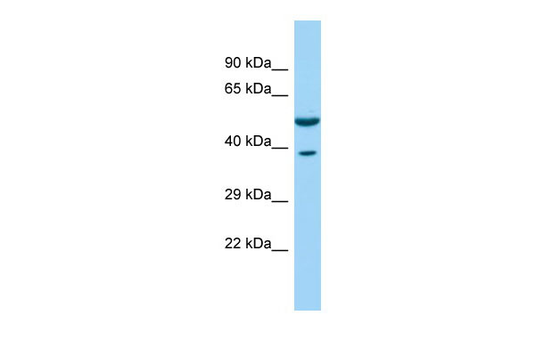
CORO1C Antibody - C-terminal region
Rabbit Polyclonal Antibody
- SPECIFICATION
- CITATIONS
- PROTOCOLS
- BACKGROUND

Application
| WB |
|---|---|
| Primary Accession | Q9ULV4 |
| Other Accession | NM_014325, NP_055140 |
| Reactivity | Human, Mouse, Rat, Rabbit, Horse, Bovine, Guinea Pig, Dog |
| Predicted | Human, Mouse, Rat, Rabbit, Pig, Horse, Bovine, Guinea Pig, Dog |
| Host | Rabbit |
| Clonality | Polyclonal |
| Calculated MW | 53kDa |
| Gene ID | 23603 |
|---|---|
| Alias Symbol | HCRNN4 |
| Other Names | Coronin-1C, Coronin-3, hCRNN4, CORO1C, CRN2, CRNN4 |
| Format | Liquid. Purified antibody supplied in 1x PBS buffer with 0.09% (w/v) sodium azide and 2% sucrose. |
| Reconstitution & Storage | Add 50 ul of distilled water. Final anti-CORO1C antibody concentration is 1 mg/ml in PBS buffer with 2% sucrose. For longer periods of storage, store at 20°C. Avoid repeat freeze-thaw cycles. |
| Precautions | CORO1C Antibody - C-terminal region is for research use only and not for use in diagnostic or therapeutic procedures. |
| Name | CORO1C {ECO:0000303|PubMed:10828594, ECO:0000312|HGNC:HGNC:2254} |
|---|---|
| Function | Plays a role in directed cell migration by regulating the activation and subcellular location of RAC1 (PubMed:25074804, PubMed:25925950). Increases the presence of activated RAC1 at the leading edge of migrating cells (PubMed:25074804, PubMed:25925950). Required for normal organization of the cytoskeleton, including the actin cytoskeleton, microtubules and the vimentin intermediate filaments (By similarity). Plays a role in endoplasmic reticulum- associated endosome fission: localizes to endosome membrane tubules and promotes recruitment of TMCC1, leading to recruitment of the endoplasmic reticulum to endosome tubules for fission (PubMed:30220460). Endosome membrane fission of early and late endosomes is essential to separate regions destined for lysosomal degradation from carriers to be recycled to the plasma membrane (PubMed:30220460). Required for normal cell proliferation, cell migration, and normal formation of lamellipodia (By similarity). Required for normal distribution of mitochondria within cells (By similarity). |
| Cellular Location | Cell membrane; Peripheral membrane protein; Cytoplasmic side. Cell projection, lamellipodium. Cell projection, ruffle membrane. Cytoplasm, cytoskeleton. Cytoplasm, cell cortex Endosome membrane. Note=All isoforms colocalize with the actin cytoskeleton in the cytosol, and especially in the cell cortex (PubMed:10828594, PubMed:19651142, PubMed:25074804) Colocalizes with F-actin at the leading edge of lamellipodia. Partially colocalizes with microtubules and vimentin intermediate filaments (PubMed:10828594, PubMed:19651142, PubMed:25074804). Localizes to endosome membrane tubules/buds (PubMed:30220460) |
| Tissue Location | Ubiquitous.. |

Thousands of laboratories across the world have published research that depended on the performance of antibodies from Abcepta to advance their research. Check out links to articles that cite our products in major peer-reviewed journals, organized by research category.
info@abcepta.com, and receive a free "I Love Antibodies" mug.

Provided below are standard protocols that you may find useful for product applications.
References
Iizaka M.,et al.Cytogenet. Cell Genet. 88:221-224(2000).
Xavier C.P.,et al.J. Mol. Biol. 393:287-299(2009).
Ota T.,et al.Nat. Genet. 36:40-45(2004).
Bechtel S.,et al.BMC Genomics 8:399-399(2007).
Scherer S.E.,et al.Nature 440:346-351(2006).
If you have used an Abcepta product and would like to share how it has performed, please click on the "Submit Review" button and provide the requested information. Our staff will examine and post your review and contact you if needed.
If you have any additional inquiries please email technical services at tech@abcepta.com.
Ordering Information
Other Products
Shipping Information
