


 Foundational characteristics of cancer include proliferation, angiogenesis, migration, evasion of apoptosis, and cellular immortality. Find key markers for these cellular processes and antibodies to detect them.
 The SUMOplot™ Analysis Program predicts and scores sumoylation sites in your protein. SUMOylation is a post-translational modification involved in various cellular processes, such as nuclear-cytosolic transport, transcriptional regulation, apoptosis, protein stability, response to stress, and progression through the cell cycle.
 The Autophagy Receptor Motif Plotter predicts and scores autophagy receptor binding sites in your protein. Identifying proteins connected to this pathway is critical to understanding the role of autophagy in physiological as well as pathological processes such as development, differentiation, neurodegenerative diseases, stress, infection, and cancer.
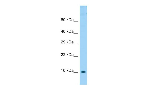
COX6B1 Antibody - N-terminal region
Rabbit Polyclonal Antibody
- SPECIFICATION
- CITATIONS
- PROTOCOLS
- BACKGROUND

Application
| WB |
|---|---|
| Primary Accession | P14854 |
| Other Accession | NM_001863, NP_001854 |
| Reactivity | Human, Mouse, Rat, Rabbit, Pig, Horse, Yeast, Bovine, Guinea Pig, Dog |
| Predicted | Human, Mouse, Rat, Rabbit, Pig, Horse, Yeast, Bovine, Guinea Pig, Dog |
| Host | Rabbit |
| Clonality | Polyclonal |
| Calculated MW | 10kDa |
| Gene ID | 1340 |
|---|---|
| Alias Symbol | COX6B, COXG, COXVIb1 |
| Other Names | Cytochrome c oxidase subunit 6B1, Cytochrome c oxidase subunit VIb isoform 1, COX VIb-1, COX6B1, COX6B |
| Format | Liquid. Purified antibody supplied in 1x PBS buffer with 0.09% (w/v) sodium azide and 2% sucrose. |
| Reconstitution & Storage | Add 50 ul of distilled water. Final anti-COX6B1 antibody concentration is 1 mg/ml in PBS buffer with 2% sucrose. For longer periods of storage, store at 20°C. Avoid repeat freeze-thaw cycles. |
| Precautions | COX6B1 Antibody - N-terminal region is for research use only and not for use in diagnostic or therapeutic procedures. |
| Name | COX6B1 |
|---|---|
| Synonyms | COX6B |
| Function | Component of the cytochrome c oxidase, the last enzyme in the mitochondrial electron transport chain which drives oxidative phosphorylation. The respiratory chain contains 3 multisubunit complexes succinate dehydrogenase (complex II, CII), ubiquinol- cytochrome c oxidoreductase (cytochrome b-c1 complex, complex III, CIII) and cytochrome c oxidase (complex IV, CIV), that cooperate to transfer electrons derived from NADH and succinate to molecular oxygen, creating an electrochemical gradient over the inner membrane that drives transmembrane transport and the ATP synthase. Cytochrome c oxidase is the component of the respiratory chain that catalyzes the reduction of oxygen to water. Electrons originating from reduced cytochrome c in the intermembrane space (IMS) are transferred via the dinuclear copper A center (CU(A)) of subunit 2 and heme A of subunit 1 to the active site in subunit 1, a binuclear center (BNC) formed by heme A3 and copper B (CU(B)). The BNC reduces molecular oxygen to 2 water molecules using 4 electrons from cytochrome c in the IMS and 4 protons from the mitochondrial matrix. |
| Cellular Location | Mitochondrion inner membrane; Peripheral membrane protein; Intermembrane side |

Thousands of laboratories across the world have published research that depended on the performance of antibodies from Abcepta to advance their research. Check out links to articles that cite our products in major peer-reviewed journals, organized by research category.
info@abcepta.com, and receive a free "I Love Antibodies" mug.

Provided below are standard protocols that you may find useful for product applications.
References
Taanman J.-W.,et al.Nucleic Acids Res. 17:1766-1766(1989).
Taanman J.-W.,et al.Gene 93:285-291(1990).
Carrero-Valenzuela R.D.,et al.Gene 102:229-236(1991).
Ota T.,et al.Nat. Genet. 36:40-45(2004).
Kalnine N.,et al.Submitted (MAY-2003) to the EMBL/GenBank/DDBJ databases.
If you have used an Abcepta product and would like to share how it has performed, please click on the "Submit Review" button and provide the requested information. Our staff will examine and post your review and contact you if needed.
If you have any additional inquiries please email technical services at tech@abcepta.com.
Ordering Information
Other Products
Shipping Information
