


 Foundational characteristics of cancer include proliferation, angiogenesis, migration, evasion of apoptosis, and cellular immortality. Find key markers for these cellular processes and antibodies to detect them.
 The SUMOplot™ Analysis Program predicts and scores sumoylation sites in your protein. SUMOylation is a post-translational modification involved in various cellular processes, such as nuclear-cytosolic transport, transcriptional regulation, apoptosis, protein stability, response to stress, and progression through the cell cycle.
 The Autophagy Receptor Motif Plotter predicts and scores autophagy receptor binding sites in your protein. Identifying proteins connected to this pathway is critical to understanding the role of autophagy in physiological as well as pathological processes such as development, differentiation, neurodegenerative diseases, stress, infection, and cancer.
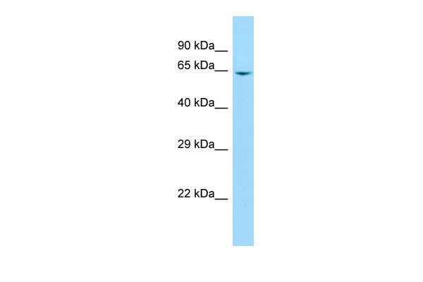
B4GALNT2 Antibody - N-terminal region
Rabbit Polyclonal Antibody
- SPECIFICATION
- CITATIONS
- PROTOCOLS
- BACKGROUND

Application
| WB |
|---|---|
| Primary Accession | Q8NHY0 |
| Other Accession | NM_153446, NP_703147 |
| Reactivity | Human |
| Predicted | Human |
| Host | Rabbit |
| Clonality | Polyclonal |
| Calculated MW | 62kDa |
| Gene ID | 124872 |
|---|---|
| Alias Symbol | B4GALT, GALGT2, MGC142235, MGC142237 |
| Other Names | Beta-1, 4 N-acetylgalactosaminyltransferase 2, 2.4.1.-, Sd(a) beta-1, 4-GalNAc transferase, UDP-GalNAc:Neu5Aca2-3Galb-R b1, 4-N-acetylgalactosaminyltransferase, B4GALNT2, GALGT2 |
| Format | Liquid. Purified antibody supplied in 1x PBS buffer with 0.09% (w/v) sodium azide and 2% sucrose. |
| Reconstitution & Storage | Add 50 ul of distilled water. Final anti-B4GALNT2 antibody concentration is 1 mg/ml in PBS buffer with 2% sucrose. For longer periods of storage, store at 20°C. Avoid repeat freeze-thaw cycles. |
| Precautions | B4GALNT2 Antibody - N-terminal region is for research use only and not for use in diagnostic or therapeutic procedures. |
| Name | B4GALNT2 {ECO:0000303|PubMed:30067891, ECO:0000312|HGNC:HGNC:24136} |
|---|---|
| Function | Beta-1,4 N-acetylgalactosaminyltransferase involved in the biosynthesis of Sd(a) histo-blood group antigen. Catalyzes the transfer of N-acetylgalactosamine (GalNAc) group in a beta-1,4-linkage from UDP- GalNAc to the galactose residue of NeuAcalpha2->3Gal-R to form Sd(a) glycan epitope GalNAcbeta1->4(NeuAcalpha2->3)Gal-R. The Sd(a) epitope is carried in O- and N-linked glycoproteins and glycolipids, including O-linked core 1 structures on GYPA/glycophorin, SLC4A1 and SLC29A1 in erythrocytes, N-linked glycans attached to the Tamm-Horsfall glycoprotein UMOD/uromodulin in renal fluids, O-linked core 3 glycans on mucins in colon and neolactosides in gastric mucosa (PubMed:12678917, PubMed:14688233, PubMed:16024623, PubMed:35409292). Confers protection against influenza A virus strains that attach to NeuAcalpha2->3-carrying host receptors. Modifies N-glycan chains on host receptors and prevents virus entry into cells (PubMed:28813663). |
| Cellular Location | [Isoform 1]: Golgi apparatus, trans-Golgi network membrane; Single-pass type II membrane protein. Cytoplasmic vesicle membrane; Single-pass type II membrane protein. Note=Partially colocalizes with EEA1 and LAMP2 in early endosomes and lysosomes, respectively. |
| Tissue Location | Widely expressed. Highly expressed in colon and to a lesser extent in kidney, stomach, ileum and rectum |

Thousands of laboratories across the world have published research that depended on the performance of antibodies from Abcepta to advance their research. Check out links to articles that cite our products in major peer-reviewed journals, organized by research category.
info@abcepta.com, and receive a free "I Love Antibodies" mug.

Provided below are standard protocols that you may find useful for product applications.
References
Montiel M.D.,et al.Biochem. J. 373:369-379(2003).
Lo Presti L.,et al.J. Biochem. 134:675-682(2003).
Ota T.,et al.Nat. Genet. 36:40-45(2004).
Zody M.C.,et al.Nature 440:1045-1049(2006).
Mural R.J.,et al.Submitted (SEP-2005) to the EMBL/GenBank/DDBJ databases.
If you have used an Abcepta product and would like to share how it has performed, please click on the "Submit Review" button and provide the requested information. Our staff will examine and post your review and contact you if needed.
If you have any additional inquiries please email technical services at tech@abcepta.com.
Ordering Information
Other Products
Shipping Information
