


 Foundational characteristics of cancer include proliferation, angiogenesis, migration, evasion of apoptosis, and cellular immortality. Find key markers for these cellular processes and antibodies to detect them.
 The SUMOplot™ Analysis Program predicts and scores sumoylation sites in your protein. SUMOylation is a post-translational modification involved in various cellular processes, such as nuclear-cytosolic transport, transcriptional regulation, apoptosis, protein stability, response to stress, and progression through the cell cycle.
 The Autophagy Receptor Motif Plotter predicts and scores autophagy receptor binding sites in your protein. Identifying proteins connected to this pathway is critical to understanding the role of autophagy in physiological as well as pathological processes such as development, differentiation, neurodegenerative diseases, stress, infection, and cancer.
MYSM1 Antibody - C-terminal region
Rabbit Polyclonal Antibody
- SPECIFICATION
- CITATIONS
- PROTOCOLS
- BACKGROUND

Application
| WB |
|---|---|
| Primary Accession | Q5VVJ2 |
| Other Accession | NM_001085487, NP_001078956 |
| Reactivity | Human, Rabbit, Dog |
| Predicted | Human, Rabbit, Dog |
| Host | Rabbit |
| Clonality | Polyclonal |
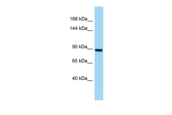
| Calculated MW | 91kDa |
| Gene ID | 114803 |
|---|---|
| Alias Symbol | 2A-DUB, 2ADUB, DKFZp779J1554, DKFZp779J1721, KIAA1915, RP4-592A1.1 |
| Other Names | Histone H2A deubiquitinase MYSM1, 2A-DUB, 3.4.19.-, Myb-like, SWIRM and MPN domain-containing protein 1, MYSM1, KIAA1915 |
| Format | Liquid. Purified antibody supplied in 1x PBS buffer with 0.09% (w/v) sodium azide and 2% sucrose. |
| Reconstitution & Storage | Add 50 ul of distilled water. Final anti-MYSM1 antibody concentration is 1 mg/ml in PBS buffer with 2% sucrose. For longer periods of storage, store at 20°C. Avoid repeat freeze-thaw cycles. |
| Precautions | MYSM1 Antibody - C-terminal region is for research use only and not for use in diagnostic or therapeutic procedures. |
| Name | MYSM1 |
|---|---|
| Synonyms | KIAA1915 |
| Function | Metalloprotease with deubiquitinase activity that plays important regulator roles in hematopoietic stem cell function, blood cell production and immune response (PubMed:24062447, PubMed:26220525, PubMed:28115216). Participates in the normal programming of B-cell responses to antigen after the maturation process (By similarity). Within the cytoplasm, plays critical roles in the repression of innate immunity and autoimmunity (PubMed:33086059). Removes 'Lys-63'-linked polyubiquitins from TRAF3 and TRAF6 complexes (By similarity). Attenuates NOD2-mediated inflammation and tissue injury by promoting 'Lys-63'-linked deubiquitination of RIPK2 component (By similarity). Suppresses the CGAS-STING1 signaling pathway by cleaving STING1 'Lys- 63'-linked ubiquitin chains (PubMed:33086059). In the nucleus, acts as a hematopoietic transcription regulator derepressing a range of genes essential for normal stem cell differentiation including EBF1 and PAX5 in B-cells, ID2 in NK-cell progenitor or FLT3 in dendritic cell precursors (PubMed:24062447). Deubiquitinates monoubiquitinated histone H2A, a specific tag for epigenetic transcriptional repression, leading to dissociation of histone H1 from the nucleosome (PubMed:17707232). |
| Cellular Location | Nucleus {ECO:0000255|PROSITE-ProRule:PRU00624, ECO:0000269|PubMed:17707232}. Cytoplasm {ECO:0000250|UniProtKB:Q69Z66} Note=Localizes to the cytoplasm in response to bacterial infection {ECO:0000250|UniProtKB:Q69Z66} |

Thousands of laboratories across the world have published research that depended on the performance of antibodies from Abcepta to advance their research. Check out links to articles that cite our products in major peer-reviewed journals, organized by research category.
info@abcepta.com, and receive a free "I Love Antibodies" mug.

Provided below are standard protocols that you may find useful for product applications.
References
Nagase T.,et al.DNA Res. 8:179-187(2001).
Ota T.,et al.Nat. Genet. 36:40-45(2004).
Bechtel S.,et al.BMC Genomics 8:399-399(2007).
Gregory S.G.,et al.Nature 441:315-321(2006).
Zhu P.,et al.Mol. Cell 27:609-621(2007).
If you have used an Abcepta product and would like to share how it has performed, please click on the "Submit Review" button and provide the requested information. Our staff will examine and post your review and contact you if needed.
If you have any additional inquiries please email technical services at tech@abcepta.com.
Ordering Information
Other Products
Shipping Information
