


 Foundational characteristics of cancer include proliferation, angiogenesis, migration, evasion of apoptosis, and cellular immortality. Find key markers for these cellular processes and antibodies to detect them.
 The SUMOplot™ Analysis Program predicts and scores sumoylation sites in your protein. SUMOylation is a post-translational modification involved in various cellular processes, such as nuclear-cytosolic transport, transcriptional regulation, apoptosis, protein stability, response to stress, and progression through the cell cycle.
 The Autophagy Receptor Motif Plotter predicts and scores autophagy receptor binding sites in your protein. Identifying proteins connected to this pathway is critical to understanding the role of autophagy in physiological as well as pathological processes such as development, differentiation, neurodegenerative diseases, stress, infection, and cancer.
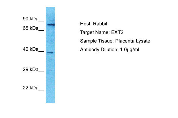
EXT2 Antibody - C-terminal region
Rabbit Polyclonal Antibody
- SPECIFICATION
- CITATIONS
- PROTOCOLS
- BACKGROUND

Application
| WB |
|---|---|
| Primary Accession | Q93063 |
| Other Accession | NP_997005 |
| Reactivity | Human |
| Host | Rabbit |
| Clonality | Polyclonal |
| Calculated MW | 78kDa |
| Gene ID | 2132 |
|---|---|
| Alias Symbol | EXT2, |
| Other Names | Exostosin-2, 2.4.1.224, 2.4.1.225, Glucuronosyl-N-acetylglucosaminyl-proteoglycan/N-acetylglucosaminyl-proteoglycan 4-alpha-N-acetylglucosaminyltransferase, Multiple exostoses protein 2, Putative tumor suppressor protein EXT2, EXT2 |
| Format | Liquid. Purified antibody supplied in 1x PBS buffer with 0.09% (w/v) sodium azide and 2% sucrose. |
| Reconstitution & Storage | Add 50 &mu, l of distilled water. Final Anti-EXT2 antibody concentration is 1 mg/ml in PBS buffer with 2% sucrose. For longer periods of storage, store at -20°C. Avoid repeat freeze-thaw cycles. |
| Precautions | EXT2 Antibody - C-terminal region is for research use only and not for use in diagnostic or therapeutic procedures. |
| Name | EXT2 (HGNC:3513) |
|---|---|
| Function | Glycosyltransferase forming with EXT1 the heterodimeric heparan sulfate polymerase which catalyzes the elongation of the heparan sulfate glycan backbone (PubMed:22660413, PubMed:36402845, PubMed:36593275). Glycan backbone extension consists in the alternating transfer of (1->4)-beta-D-GlcA and (1->4)-alpha-D-GlcNAc residues from their respective UDP-sugar donors. Both EXT1 and EXT2 are required for the full activity of the polymerase since EXT1 bears the N- acetylglucosaminyl-proteoglycan 4-beta-glucuronosyltransferase activity within the complex while EXT2 carries the glucuronosyl-N- acetylglucosaminyl-proteoglycan 4-alpha-N-acetylglucosaminyltransferase activity (PubMed:36402845, PubMed:36593275). Heparan sulfate proteoglycans are ubiquitous components of the extracellular matrix and play an important role in tissue homeostasis and signaling (PubMed:19344451, PubMed:22660413). |
| Cellular Location | Golgi apparatus membrane; Single-pass type II membrane protein. Golgi apparatus, cis-Golgi network membrane; Single-pass type II membrane protein. Endoplasmic reticulum membrane; Single-pass type II membrane protein. Secreted {ECO:0000250|UniProtKB:O77783}. Note=The active heparan sulfate polymerase complex composed of EXT1 and EXT2 is localized to the Golgi apparatus. If both proteins are individually detected in the endoplasmic reticulum, the formation of the complex promotes their transport to the Golgi. |
| Tissue Location | Widely expressed.. |

Thousands of laboratories across the world have published research that depended on the performance of antibodies from Abcepta to advance their research. Check out links to articles that cite our products in major peer-reviewed journals, organized by research category.
info@abcepta.com, and receive a free "I Love Antibodies" mug.

Provided below are standard protocols that you may find useful for product applications.
Background
Glycosyltransferase required for the biosynthesis of heparan-sulfate. The EXT1/EXT2 complex possesses substantially higher glycosyltransferase activity than EXT1 or EXT2 alone. Appears to be a tumor suppressor.
References
Stickens D.J.,et al.Nat. Genet. 14:25-32(1996).
Wuyts W.,et al.Hum. Mol. Genet. 5:1547-1557(1996).
Clines G.A.,et al.Genome Res. 7:359-367(1997).
Deng H.-X.,et al.Prog. Nat. Sci. 6:692-699(1996).
Ota T.,et al.Nat. Genet. 36:40-45(2004).
If you have used an Abcepta product and would like to share how it has performed, please click on the "Submit Review" button and provide the requested information. Our staff will examine and post your review and contact you if needed.
If you have any additional inquiries please email technical services at tech@abcepta.com.
Ordering Information
Other Products
Shipping Information
