


 Foundational characteristics of cancer include proliferation, angiogenesis, migration, evasion of apoptosis, and cellular immortality. Find key markers for these cellular processes and antibodies to detect them.
 The SUMOplot™ Analysis Program predicts and scores sumoylation sites in your protein. SUMOylation is a post-translational modification involved in various cellular processes, such as nuclear-cytosolic transport, transcriptional regulation, apoptosis, protein stability, response to stress, and progression through the cell cycle.
 The Autophagy Receptor Motif Plotter predicts and scores autophagy receptor binding sites in your protein. Identifying proteins connected to this pathway is critical to understanding the role of autophagy in physiological as well as pathological processes such as development, differentiation, neurodegenerative diseases, stress, infection, and cancer.
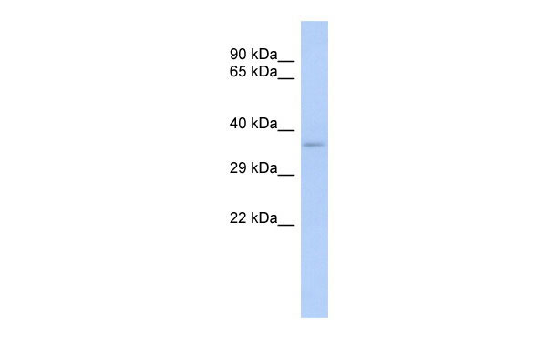
TRADD antibody - middle region
Rabbit Polyclonal Antibody
- SPECIFICATION
- CITATIONS
- PROTOCOLS
- BACKGROUND

Application
| WB |
|---|---|
| Primary Accession | Q15628 |
| Other Accession | NM_003789, NP_003780 |
| Reactivity | Human, Horse, Dog |
| Predicted | Human, Horse, Dog |
| Host | Rabbit |
| Clonality | Polyclonal |
| Calculated MW | 34kDa |
| Gene ID | 8717 |
|---|---|
| Alias Symbol | Hs.89862, MGC11078 |
| Other Names | Tumor necrosis factor receptor type 1-associated DEATH domain protein, TNFR1-associated DEATH domain protein, TNFRSF1A-associated via death domain, TRADD |
| Format | Liquid. Purified antibody supplied in 1x PBS buffer with 0.09% (w/v) sodium azide and 2% sucrose. |
| Reconstitution & Storage | Add 50 ul of distilled water. Final anti-TRADD antibody concentration is 1 mg/ml in PBS buffer with 2% sucrose. For longer periods of storage, store at 20°C. Avoid repeat freeze-thaw cycles. |
| Precautions | TRADD antibody - middle region is for research use only and not for use in diagnostic or therapeutic procedures. |
| Name | TRADD {ECO:0000303|PubMed:7758105, ECO:0000312|HGNC:HGNC:12030} |
|---|---|
| Function | Adapter molecule for TNFRSF1A/TNFR1 that specifically associates with the cytoplasmic domain of activated TNFRSF1A/TNFR1 mediating its interaction with FADD (PubMed:23955153, PubMed:7758105, PubMed:8612133). Overexpression of TRADD leads to two major TNF-induced responses, apoptosis and activation of NF-kappa-B (PubMed:7758105, PubMed:8612133). The nuclear form acts as a tumor suppressor by preventing ubiquitination and degradation of isoform p19ARF/ARF of CDKN2A by TRIP12: acts by interacting with TRIP12, leading to disrupt interaction between TRIP12 and isoform p19ARF/ARF of CDKN2A (By similarity). |
| Cellular Location | Nucleus {ECO:0000250|UniProtKB:Q3U0V2}. Cytoplasm. Cytoplasm, cytoskeleton. Note=Shuttles between the cytoplasm and the nucleus. {ECO:0000250|UniProtKB:Q3U0V2} |
| Tissue Location | Found in all examined tissues. |

Thousands of laboratories across the world have published research that depended on the performance of antibodies from Abcepta to advance their research. Check out links to articles that cite our products in major peer-reviewed journals, organized by research category.
info@abcepta.com, and receive a free "I Love Antibodies" mug.

Provided below are standard protocols that you may find useful for product applications.
Background
The nuclear form acts as a tumor suppressor by preventing ubiquitination and degradation of isoform p19ARF/ARF of CDKN2A by TRIP12: acts by interacting with TRIP12, leading to disrupt interaction between TRIP12 and isoform p19ARF/ARF of CDKN2A (By similarity). Adapter molecule for TNFRSF1A/TNFR1 that specifically associates with the cytoplasmic domain of activated TNFRSF1A/TNFR1 mediating its interaction with FADD. Overexpression of TRADD leads to two major TNF-induced responses, apoptosis and activation of NF-kappa-B.
References
Hsu H.,et al.Cell 81:495-504(1995).
Scheuerpflug C.G.,et al.Submitted (JUL-2001) to the EMBL/GenBank/DDBJ databases.
Kaiser C.,et al.Submitted (MAR-2005) to the EMBL/GenBank/DDBJ databases.
Kalnine N.,et al.Submitted (OCT-2004) to the EMBL/GenBank/DDBJ databases.
Ota T.,et al.Nat. Genet. 36:40-45(2004).
If you have used an Abcepta product and would like to share how it has performed, please click on the "Submit Review" button and provide the requested information. Our staff will examine and post your review and contact you if needed.
If you have any additional inquiries please email technical services at tech@abcepta.com.
Ordering Information
Other Products
Shipping Information
