


 Foundational characteristics of cancer include proliferation, angiogenesis, migration, evasion of apoptosis, and cellular immortality. Find key markers for these cellular processes and antibodies to detect them.
 The SUMOplot™ Analysis Program predicts and scores sumoylation sites in your protein. SUMOylation is a post-translational modification involved in various cellular processes, such as nuclear-cytosolic transport, transcriptional regulation, apoptosis, protein stability, response to stress, and progression through the cell cycle.
 The Autophagy Receptor Motif Plotter predicts and scores autophagy receptor binding sites in your protein. Identifying proteins connected to this pathway is critical to understanding the role of autophagy in physiological as well as pathological processes such as development, differentiation, neurodegenerative diseases, stress, infection, and cancer.
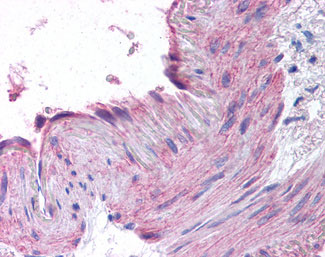
CCR1 Antibody (N-Terminus)
Rabbit Polyclonal Antibody
- SPECIFICATION
- CITATIONS
- PROTOCOLS
- BACKGROUND

Application
| IHC-P |
|---|---|
| Primary Accession | P32246 |
| Reactivity | Human |
| Host | Rabbit |
| Clonality | Polyclonal |
| Calculated MW | 41kDa |
| Dilution | IHC-P (2.5 µg/ml) |
| Gene ID | 1230 |
|---|---|
| Other Names | C-C chemokine receptor type 1, C-C CKR-1, CC-CKR-1, CCR-1, CCR1, HM145, LD78 receptor, Macrophage inflammatory protein 1-alpha receptor, MIP-1alpha-R, RANTES-R, CD191, CCR1, CMKBR1, CMKR1, SCYAR1 |
| Target/Specificity | Human CCR1. BLAST analysis of the peptide immunogen showed no homology with other human proteins. |
| Reconstitution & Storage | Long term: -70°C; Short term: +4°C |
| Precautions | CCR1 Antibody (N-Terminus) is for research use only and not for use in diagnostic or therapeutic procedures. |
| Name | CCR1 |
|---|---|
| Synonyms | CMKBR1, CMKR1, SCYAR1 |
| Function | Chemokine receptor that plays a crucial role in regulating immune cell migration, inflammation, and immune responses (PubMed:14991608). Contributes to the inflammatory response by recruiting immune cells, such as monocytes, macrophages, T-cells, and dendritic cells, to sites of inflammation for the clearance of pathogens and the resolution of tissue damage. When activated by its ligands including CCL3, CCL5-9, CCL13-16 and CCL23, triggers a signaling cascade within immune cells, leading to their migration towards the source of the chemokine (PubMed:15905581). For example, mediates neutrophil migration after activation by CCL3 leading to the sequential release of TNF-alpha and leukotriene B4 (By similarity). Also mediates monocyte migration upon CXCL4 binding (PubMed:29930254). Activation by CCL5 results in neuroinflammation through the ERK1/2 signaling pathway (By similarity). |
| Cellular Location | Cell membrane; Multi-pass membrane protein |
| Tissue Location | Widely expressed in different hematopoietic cells. |
| Volume | 62.5 µl |

Thousands of laboratories across the world have published research that depended on the performance of antibodies from Abcepta to advance their research. Check out links to articles that cite our products in major peer-reviewed journals, organized by research category.
info@abcepta.com, and receive a free "I Love Antibodies" mug.

Provided below are standard protocols that you may find useful for product applications.
Background
Receptor for a C-C type chemokine. Binds to MIP-1-alpha, MIP-1-delta, RANTES, and MCP-3 and, less efficiently, to MIP-1- beta or MCP-1 and subsequently transduces a signal by increasing the intracellular calcium ions level. Responsible for affecting stem cell proliferation.
References
Neote K.,et al.Cell 72:415-425(1993).
Gao J.-L.,et al.J. Exp. Med. 177:1421-1427(1993).
Nomura H.,et al.Int. Immunol. 5:1239-1249(1993).
Ko J.,et al.FASEB J. 18:890-892(2004).
Sung H.J.,et al.Exp. Mol. Med. 40:332-338(2008).
If you have used an Abcepta product and would like to share how it has performed, please click on the "Submit Review" button and provide the requested information. Our staff will examine and post your review and contact you if needed.
If you have any additional inquiries please email technical services at tech@abcepta.com.
Ordering Information
Other Products
Shipping Information
