


 Foundational characteristics of cancer include proliferation, angiogenesis, migration, evasion of apoptosis, and cellular immortality. Find key markers for these cellular processes and antibodies to detect them.
 The SUMOplot™ Analysis Program predicts and scores sumoylation sites in your protein. SUMOylation is a post-translational modification involved in various cellular processes, such as nuclear-cytosolic transport, transcriptional regulation, apoptosis, protein stability, response to stress, and progression through the cell cycle.
 The Autophagy Receptor Motif Plotter predicts and scores autophagy receptor binding sites in your protein. Identifying proteins connected to this pathway is critical to understanding the role of autophagy in physiological as well as pathological processes such as development, differentiation, neurodegenerative diseases, stress, infection, and cancer.
GPRC5A / RAI3 Antibody (Cytoplasmic Domain)
Rabbit Polyclonal Antibody
- SPECIFICATION
- CITATIONS
- PROTOCOLS
- BACKGROUND

Application
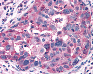
| IHC-P |
|---|---|
| Primary Accession | Q8NFJ5 |
| Reactivity | Human |
| Host | Rabbit |
| Clonality | Polyclonal |
| Calculated MW | 40kDa |
| Dilution | IHC-P (5-6 µg/ml) |
| Gene ID | 9052 |
|---|---|
| Other Names | Retinoic acid-induced protein 3, G-protein coupled receptor family C group 5 member A, Orphan G-protein-coupling receptor PEIG-1, Retinoic acid-induced gene 1 protein, RAIG-1, GPRC5A, GPCR5A, RAI3, RAIG1 |
| Target/Specificity | Human GPRC5A / RAI3. BLAST analysis of the peptide immunogen showed no homology with other human proteins. |
| Reconstitution & Storage | Long term: -70°C; Short term: +4°C |
| Precautions | GPRC5A / RAI3 Antibody (Cytoplasmic Domain) is for research use only and not for use in diagnostic or therapeutic procedures. |
| Name | GPRC5A |
|---|---|
| Synonyms | GPCR5A, RAI3, RAIG1 |
| Function | Orphan receptor. Could be involved in modulating differentiation and maintaining homeostasis of epithelial cells. This retinoic acid-inducible GPCR provide evidence for a possible interaction between retinoid and G-protein signaling pathways. Functions as a negative modulator of EGFR signaling (By similarity). May act as a lung tumor suppressor (PubMed:18000218). |
| Cellular Location | Cell membrane; Multi-pass membrane protein. Cytoplasmic vesicle membrane; Multi-pass membrane protein. Note=Localized in perinuclear vesicles, probably Golgi- associated vesicles. |
| Tissue Location | Expressed at high level in fetal and adult lung tissues but repressed in most human lung cancers (PubMed:18000218, PubMed:9857033). Constitutively expressed in fetal kidney and adult placenta, kidney, prostate, testis, ovary, small intestine, colon, stomach, and spinal cord at low to moderate levels. Not detectable in fetal heart, brain, and liver and adult heart, brain, liver, skeletal muscle, pancreas, spleen, thymus, and peripheral leukocytes. According to PubMed:10783259, expressed at low but detectable level in pancreas and heart. |
| Volume | 50 µl |

Thousands of laboratories across the world have published research that depended on the performance of antibodies from Abcepta to advance their research. Check out links to articles that cite our products in major peer-reviewed journals, organized by research category.
info@abcepta.com, and receive a free "I Love Antibodies" mug.

Provided below are standard protocols that you may find useful for product applications.
Background
Unknown. This G-protein coupled receptor could be involved in modulating differentiation and maintaining homeostasis of epithelial cells. The comparable expression level in fetal lung and kidney with adult tissues suggests a possible role in embryonic development and maturation of these organs. This retinoic acid-inducible GPCR provide evidence for a possible interaction between retinoid and G-protein signaling pathways.
References
Cheng Y.,et al.J. Biol. Chem. 273:35008-35015(1998).
Cafferata E.G.,et al.Submitted (APR-2002) to the EMBL/GenBank/DDBJ databases.
Ota T.,et al.Nat. Genet. 36:40-45(2004).
Mural R.J.,et al.Submitted (JUL-2005) to the EMBL/GenBank/DDBJ databases.
Braeuner-Osborne H.,et al.Genomics 65:121-128(2000).
If you have used an Abcepta product and would like to share how it has performed, please click on the "Submit Review" button and provide the requested information. Our staff will examine and post your review and contact you if needed.
If you have any additional inquiries please email technical services at tech@abcepta.com.
Ordering Information
Other Products
Shipping Information
