


 Foundational characteristics of cancer include proliferation, angiogenesis, migration, evasion of apoptosis, and cellular immortality. Find key markers for these cellular processes and antibodies to detect them.
 The SUMOplot™ Analysis Program predicts and scores sumoylation sites in your protein. SUMOylation is a post-translational modification involved in various cellular processes, such as nuclear-cytosolic transport, transcriptional regulation, apoptosis, protein stability, response to stress, and progression through the cell cycle.
 The Autophagy Receptor Motif Plotter predicts and scores autophagy receptor binding sites in your protein. Identifying proteins connected to this pathway is critical to understanding the role of autophagy in physiological as well as pathological processes such as development, differentiation, neurodegenerative diseases, stress, infection, and cancer.
ADORA2A/Adenosine A2A Receptor Antibody (C-Terminus)
Rabbit Polyclonal Antibody
- SPECIFICATION
- CITATIONS
- PROTOCOLS
- BACKGROUND

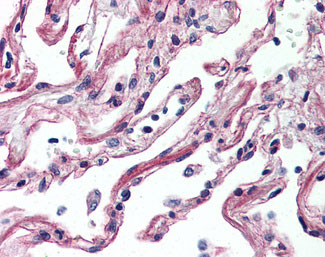
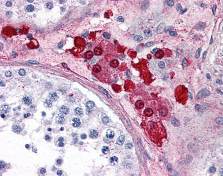
Application
| IHC-P |
|---|---|
| Primary Accession | P29274 |
| Reactivity | Human |
| Host | Rabbit |
| Clonality | Polyclonal |
| Calculated MW | 45kDa |
| Dilution | IHC-P (10 µg/ml) |
| Gene ID | 135 |
|---|---|
| Other Names | Adenosine receptor A2a, ADORA2A, ADORA2 |
| Target/Specificity | Human ADORA2A. BLAST analysis of the peptide immunogen showed no homology with other human proteins. |
| Reconstitution & Storage | Long term: -70°C; Short term: +4°C |
| Precautions | ADORA2A/Adenosine A2A Receptor Antibody (C-Terminus) is for research use only and not for use in diagnostic or therapeutic procedures. |
| Name | ADORA2A |
|---|---|
| Synonyms | ADORA2 |
| Function | Receptor for adenosine (By similarity). The activity of this receptor is mediated by G proteins which activate adenylyl cyclase (By similarity). |
| Cellular Location | Cell membrane {ECO:0000250|UniProtKB:P30543}; Multi-pass membrane protein {ECO:0000250|UniProtKB:P30543} Note=Colocalizes with GAS2L2 at neuronal processes {ECO:0000250|UniProtKB:P30543} |

Thousands of laboratories across the world have published research that depended on the performance of antibodies from Abcepta to advance their research. Check out links to articles that cite our products in major peer-reviewed journals, organized by research category.
info@abcepta.com, and receive a free "I Love Antibodies" mug.

Provided below are standard protocols that you may find useful for product applications.
Background
Receptor for adenosine. The activity of this receptor is mediated by G proteins which activate adenylyl cyclase.
References
Tiffany H.L.,et al.Submitted (JUL-1992) to the EMBL/GenBank/DDBJ databases.
Salvatore C.A.,et al.Submitted (SEP-1992) to the EMBL/GenBank/DDBJ databases.
Furlong T.J.,et al.Brain Res. Mol. Brain Res. 15:62-66(1992).
Le F.,et al.Biochem. Biophys. Res. Commun. 223:461-467(1996).
Puhl H.L. III,et al.Submitted (JUL-2002) to the EMBL/GenBank/DDBJ databases.
If you have used an Abcepta product and would like to share how it has performed, please click on the "Submit Review" button and provide the requested information. Our staff will examine and post your review and contact you if needed.
If you have any additional inquiries please email technical services at tech@abcepta.com.
Ordering Information
Other Products
Shipping Information
