


 Foundational characteristics of cancer include proliferation, angiogenesis, migration, evasion of apoptosis, and cellular immortality. Find key markers for these cellular processes and antibodies to detect them.
 The SUMOplot™ Analysis Program predicts and scores sumoylation sites in your protein. SUMOylation is a post-translational modification involved in various cellular processes, such as nuclear-cytosolic transport, transcriptional regulation, apoptosis, protein stability, response to stress, and progression through the cell cycle.
 The Autophagy Receptor Motif Plotter predicts and scores autophagy receptor binding sites in your protein. Identifying proteins connected to this pathway is critical to understanding the role of autophagy in physiological as well as pathological processes such as development, differentiation, neurodegenerative diseases, stress, infection, and cancer.
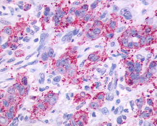
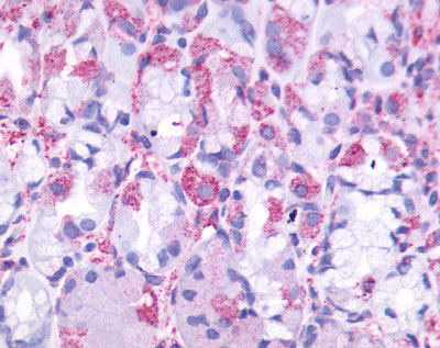
CCKBR / Cckb Antibody (Cytoplasmic Domain)
Rabbit Polyclonal Antibody
- SPECIFICATION
- CITATIONS
- PROTOCOLS
- BACKGROUND

Application
| IHC-P |
|---|---|
| Primary Accession | P32239 |
| Reactivity | Human |
| Host | Rabbit |
| Clonality | Polyclonal |
| Calculated MW | 48kDa |
| Dilution | IHC-P (3 µg/ml) |
| Gene ID | 887 |
|---|---|
| Other Names | Gastrin/cholecystokinin type B receptor, CCK-B receptor, CCK-BR, Cholecystokinin-2 receptor, CCK2-R, CCKBR, CCKRB |
| Target/Specificity | Human CCKBR. BLAST analysis of the peptide immunogen showed no homology with other human proteins. |
| Reconstitution & Storage | Long term: -70°C; Short term: +4°C |
| Precautions | CCKBR / Cckb Antibody (Cytoplasmic Domain) is for research use only and not for use in diagnostic or therapeutic procedures. |
| Name | CCKBR (HGNC:1571) |
|---|---|
| Synonyms | CCKRB |
| Function | Receptor for the peptide hormones gastrin and cholecystokinin (CCK). Expressed throughout the central nervous system, where it modulates processes such as anxiety, analgesia, arousal and neuroleptic activity. Couples to both GNAI1 and GNAQ signaling pathways, but not to GNAS (PubMed:34556863). Upon gastrin activation, reduces glucose absorption in intestinal epithelial cells by downregulating SGLT1 and GLUT2 expression through suppression of the PI3K/Akt/eIF4B pathway (By similarity). In the kidney, decreases SGLT2 expression under high- glucose conditions via ERK/NF-kappa-B signaling (By similarity). |
| Cellular Location | Cell membrane; Multi-pass membrane protein. |
| Tissue Location | Isoform 1 is expressed in brain, pancreas, stomach, the colon cancer cell line LoVo and the T-lymphoblastoma Jurkat, but not in heart, placenta, liver, lung, skeletal muscle, kidney or the stomach cancer cell line AGS. Expressed at high levels in the small cell lung cancer cell line NCI-H510, at lower levels in NCI-H345, NCI- H69 and GLC-28 cell lines, not expressed in GLC-19 cell line. Within the stomach, expressed at high levels in the mucosa of the gastric fundus and at low levels in the antrum and duodenum. Isoform 2 is present in pancreatic cancer cells and colorectal cancer cells, but not in normal pancreas or colonic mucosa. Isoform 3 is expressed in brain, pancreas, stomach, the stomach cancer cell line AGS and the colon cancer cell line LoVo. |
| Volume | 50 µl |

Thousands of laboratories across the world have published research that depended on the performance of antibodies from Abcepta to advance their research. Check out links to articles that cite our products in major peer-reviewed journals, organized by research category.
info@abcepta.com, and receive a free "I Love Antibodies" mug.

Provided below are standard protocols that you may find useful for product applications.
Background
Receptor for gastrin and cholecystokinin. The CKK-B receptors occur throughout the central nervous system where they modulate anxiety, analgesia, arousal, and neuroleptic activity. This receptor mediates its action by association with G proteins that activate a phosphatidylinositol-calcium second messenger system.
References
Pisegna J.R.,et al.Biochem. Biophys. Res. Commun. 189:296-303(1992).
Lee Y.-M.,et al.J. Biol. Chem. 268:8164-8169(1993).
Ito M.,et al.J. Biol. Chem. 268:18300-18305(1993).
Song I.,et al.Proc. Natl. Acad. Sci. U.S.A. 90:9085-9089(1993).
Herget T.,et al.Ann. N. Y. Acad. Sci. 713:283-297(1994).
If you have used an Abcepta product and would like to share how it has performed, please click on the "Submit Review" button and provide the requested information. Our staff will examine and post your review and contact you if needed.
If you have any additional inquiries please email technical services at tech@abcepta.com.
Ordering Information
Other Products
Shipping Information
