


 Foundational characteristics of cancer include proliferation, angiogenesis, migration, evasion of apoptosis, and cellular immortality. Find key markers for these cellular processes and antibodies to detect them.
 The SUMOplot™ Analysis Program predicts and scores sumoylation sites in your protein. SUMOylation is a post-translational modification involved in various cellular processes, such as nuclear-cytosolic transport, transcriptional regulation, apoptosis, protein stability, response to stress, and progression through the cell cycle.
 The Autophagy Receptor Motif Plotter predicts and scores autophagy receptor binding sites in your protein. Identifying proteins connected to this pathway is critical to understanding the role of autophagy in physiological as well as pathological processes such as development, differentiation, neurodegenerative diseases, stress, infection, and cancer.
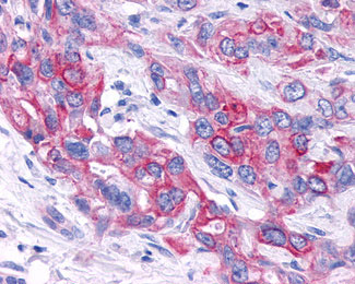
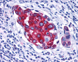
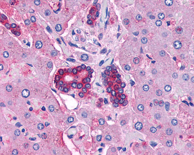
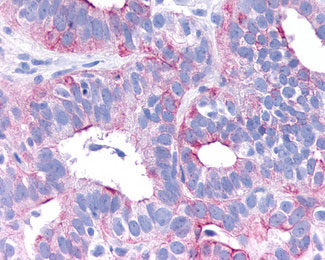
VPAC2 / VIPR2 Antibody (C-Terminus)
Rabbit Polyclonal Antibody
- SPECIFICATION
- CITATIONS
- PROTOCOLS
- BACKGROUND

Application
| IHC-P |
|---|---|
| Primary Accession | P41587 |
| Reactivity | Human |
| Host | Rabbit |
| Clonality | Polyclonal |
| Calculated MW | 49kDa |
| Dilution | IHC-P (3-5 µg/ml) |
| Gene ID | 7434 |
|---|---|
| Other Names | Vasoactive intestinal polypeptide receptor 2, VIP-R-2, Helodermin-preferring VIP receptor, Pituitary adenylate cyclase-activating polypeptide type III receptor, PACAP type III receptor, PACAP-R-3, PACAP-R3, VPAC2, VIPR2, VIP2R |
| Target/Specificity | Human VIPR2. BLAST analysis of the peptide immunogen showed no homology with other human proteins. |
| Reconstitution & Storage | Long term: -70°C; Short term: +4°C |
| Precautions | VPAC2 / VIPR2 Antibody (C-Terminus) is for research use only and not for use in diagnostic or therapeutic procedures. |
| Name | VIPR2 (HGNC:12695) |
|---|---|
| Synonyms | VIP2R |
| Function | G protein-coupled receptor activated by the neuropeptides vasoactive intestinal peptide (VIP) and pituitary adenylate cyclase- activating polypeptide (ADCYAP1/PACAP) (PubMed:7811244, PubMed:35477937, PubMed:8933357). Binds VIP and both PACAP27 and PACAP38 bioactive peptides with the following order of potency PACAP38 = VIP > PACAP27 (PubMed:35477937, PubMed:8933357). Ligand binding causes a conformation change that triggers signaling via guanine nucleotide-binding proteins (G proteins) and modulates the activity of downstream effectors. Activates cAMP-dependent pathway (PubMed:7811244, PubMed:35477937, PubMed:8933357). May be coupled to phospholipase C. |
| Cellular Location | Cell membrane; Multi-pass membrane protein |
| Tissue Location | Expressed in CD4+ T-cells, but not in CD8+ T-cells. Expressed in the T-cell lines Jurkat, Peer, MOLT-4, HSB, YT and SUP-T1, but not in the T-cell lines HARRIS and HuT 78 |
| Volume | 50 µl |

Thousands of laboratories across the world have published research that depended on the performance of antibodies from Abcepta to advance their research. Check out links to articles that cite our products in major peer-reviewed journals, organized by research category.
info@abcepta.com, and receive a free "I Love Antibodies" mug.

Provided below are standard protocols that you may find useful for product applications.
Background
This is a receptor for VIP as well as PACAP-38 and -27, the activity of this receptor is mediated by G proteins which activate adenylyl cyclase. Can be coupled to phospholipase C.
References
Svoboda M.,et al.Biochem. Biophys. Res. Commun. 205:1617-1624(1994).
Mackay M.,et al.Genomics 37:345-353(1996).
Wei Y.,et al.J. Neuroendocrinol. 8:811-817(1996).
Lutz E.M.,et al.FEBS Lett. 458:197-203(1999).
Kalnine N.,et al.Submitted (MAY-2003) to the EMBL/GenBank/DDBJ databases.
If you have used an Abcepta product and would like to share how it has performed, please click on the "Submit Review" button and provide the requested information. Our staff will examine and post your review and contact you if needed.
If you have any additional inquiries please email technical services at tech@abcepta.com.
Ordering Information
Other Products
Shipping Information
