


 Foundational characteristics of cancer include proliferation, angiogenesis, migration, evasion of apoptosis, and cellular immortality. Find key markers for these cellular processes and antibodies to detect them.
 The SUMOplot™ Analysis Program predicts and scores sumoylation sites in your protein. SUMOylation is a post-translational modification involved in various cellular processes, such as nuclear-cytosolic transport, transcriptional regulation, apoptosis, protein stability, response to stress, and progression through the cell cycle.
 The Autophagy Receptor Motif Plotter predicts and scores autophagy receptor binding sites in your protein. Identifying proteins connected to this pathway is critical to understanding the role of autophagy in physiological as well as pathological processes such as development, differentiation, neurodegenerative diseases, stress, infection, and cancer.
CCR3 Antibody (Extracellular Domain)
Rabbit Polyclonal Antibody
- SPECIFICATION
- CITATIONS
- PROTOCOLS
- BACKGROUND

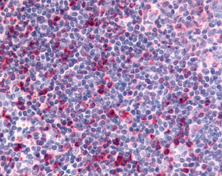
Application
| IHC-P, E |
|---|---|
| Primary Accession | P51677 |
| Reactivity | Human |
| Host | Rabbit |
| Clonality | Polyclonal |
| Calculated MW | 41kDa |
| Dilution | IHC-P (10 µg/ml) |
| Gene ID | 1232 |
|---|---|
| Other Names | C-C chemokine receptor type 3, C-C CKR-3, CC-CKR-3, CCR-3, CCR3, CKR3, Eosinophil eotaxin receptor, CD193, CCR3, CMKBR3 |
| Target/Specificity | Human CCR3. BLAST analysis of the peptide immunogen showed no homology with other human proteins. |
| Reconstitution & Storage | Long term: -70°C; Short term: +4°C |
| Precautions | CCR3 Antibody (Extracellular Domain) is for research use only and not for use in diagnostic or therapeutic procedures. |
| Name | CCR3 |
|---|---|
| Synonyms | CMKBR3 |
| Function | G protein-coupled receptor (GPCR) that plays a key role in the immune system by regulating the migration and activation of white blood cells in response to chemokines (PubMed:28994588). Selectively interacts with eosinophil-attracting chemokines such as eotaxin/CCL11, eotaxin-2/CCL24 and eotaxin-3/CCL26 (PubMed:7622448, PubMed:8642344, PubMed:8676064). Ligand binding triggers intracellular signaling that leads to chemotaxis of immune cells. Mechanistically, signals through GNA14 or GNA16 to induce stimulation of phospholipase Cbeta/PLCB2 and subsequently chemotaxis (PubMed:18406577). Alternatively, transduces signal via GNAI1 resulting in elevated intracellular calcium levels and activation of the PI3K/AKT pathway (PubMed:8676064, PubMed:35570218). May also act as a possible functional receptor for NARS1 (PubMed:30171954). |
| Cellular Location | Cell membrane; Multi-pass membrane protein |
| Tissue Location | In eosinophils as well as trace amounts in neutrophils and monocytes. |
| Volume | 50 µl |

Thousands of laboratories across the world have published research that depended on the performance of antibodies from Abcepta to advance their research. Check out links to articles that cite our products in major peer-reviewed journals, organized by research category.
info@abcepta.com, and receive a free "I Love Antibodies" mug.

Provided below are standard protocols that you may find useful for product applications.
Background
Receptor for a C-C type chemokine. Binds to eotaxin, eotaxin-3, MCP-3, MCP-4, RANTES and MIP-1 delta. Subsequently transduces a signal by increasing the intracellular calcium ions level. Alternative coreceptor with CD4 for HIV-1 infection.
References
Combadiere C.,et al.J. Biol. Chem. 270:16491-16494(1995).
Combadiere C.,et al.J. Biol. Chem. 270:30235-30235(1995).
Daugherty B.L.,et al.J. Exp. Med. 183:2349-2354(1996).
Ponath P.D.,et al.J. Exp. Med. 183:2437-2448(1996).
Xiao L.,et al.Submitted (NOV-1997) to the EMBL/GenBank/DDBJ databases.
If you have used an Abcepta product and would like to share how it has performed, please click on the "Submit Review" button and provide the requested information. Our staff will examine and post your review and contact you if needed.
If you have any additional inquiries please email technical services at tech@abcepta.com.
Ordering Information
Other Products
Shipping Information
