


 Foundational characteristics of cancer include proliferation, angiogenesis, migration, evasion of apoptosis, and cellular immortality. Find key markers for these cellular processes and antibodies to detect them.
 The SUMOplot™ Analysis Program predicts and scores sumoylation sites in your protein. SUMOylation is a post-translational modification involved in various cellular processes, such as nuclear-cytosolic transport, transcriptional regulation, apoptosis, protein stability, response to stress, and progression through the cell cycle.
 The Autophagy Receptor Motif Plotter predicts and scores autophagy receptor binding sites in your protein. Identifying proteins connected to this pathway is critical to understanding the role of autophagy in physiological as well as pathological processes such as development, differentiation, neurodegenerative diseases, stress, infection, and cancer.
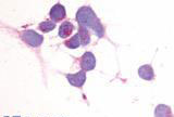
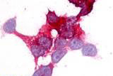
ADGRE2 / EMR2 Antibody (N-Terminus)
Rabbit Polyclonal Antibody
- SPECIFICATION
- CITATIONS
- PROTOCOLS
- BACKGROUND

Application
| IHC-P, ICC, E |
|---|---|
| Primary Accession | Q9UHX3 |
| Reactivity | Human |
| Host | Rabbit |
| Clonality | Polyclonal |
| Calculated MW | 90kDa |
| Dilution | IHC-P (7.5-15 µg/ml) |
| Gene ID | 30817 |
|---|---|
| Other Names | EGF-like module-containing mucin-like hormone receptor-like 2, EGF-like module receptor 2, CD312, EMR2 |
| Target/Specificity | Human EMR2. BLAST analysis of the peptide immunogen showed no homology with other human proteins, except MYO18B (45%), FLJ13231 (40%). |
| Reconstitution & Storage | Long term: -70°C; Short term: +4°C |
| Precautions | ADGRE2 / EMR2 Antibody (N-Terminus) is for research use only and not for use in diagnostic or therapeutic procedures. |
| Name | ADGRE2 (HGNC:3337) |
|---|---|
| Synonyms | EMR2 |
| Function | Cell surface receptor that binds to the chondroitin sulfate moiety of glycosaminoglycan chains and promotes cell attachment. Promotes granulocyte chemotaxis, degranulation and adhesion. In macrophages, promotes the release of inflammatory cytokines, including IL8 and TNF. Signals probably through G-proteins. Is a regulator of mast cell degranulation (PubMed:26841242). |
| Cellular Location | Cell membrane; Multi-pass membrane protein. Cell projection, ruffle membrane; Multi-pass membrane protein. Note=Localized at the leading edge of migrating cells. |
| Tissue Location | Expression is restricted to myeloid cells. Highest expression was found in peripheral blood leukocytes, followed by spleen and lymph nodes, with intermediate to low levels in thymus, bone marrow, fetal liver, placenta, and lung, and no expression in heart, brain, skeletal muscle, kidney, or pancreas. Expression is also detected in monocyte/macrophage and Jurkat cell lines but not in other cell lines tested. High expression in mast cells (PubMed:26841242) |
| Volume | 50 µl |

Thousands of laboratories across the world have published research that depended on the performance of antibodies from Abcepta to advance their research. Check out links to articles that cite our products in major peer-reviewed journals, organized by research category.
info@abcepta.com, and receive a free "I Love Antibodies" mug.

Provided below are standard protocols that you may find useful for product applications.
Background
Cell surface receptor that binds to the chondroitin sulfate moiety of glycosaminoglycan chains and promotes cell attachment. Promotes granulocyte chemotaxis, degranulation and adhesion. In macrophages, promotes the release of inflammatory cytokines, including IL8 and TNF.
References
Lin H.-H.,et al.Genomics 67:188-200(2000).
Suwa M.,et al.Submitted (JUL-2001) to the EMBL/GenBank/DDBJ databases.
Ota T.,et al.Nat. Genet. 36:40-45(2004).
Grimwood J.,et al.Nature 428:529-535(2004).
Chang G.-W.,et al.FEBS Lett. 547:145-150(2003).
If you have used an Abcepta product and would like to share how it has performed, please click on the "Submit Review" button and provide the requested information. Our staff will examine and post your review and contact you if needed.
If you have any additional inquiries please email technical services at tech@abcepta.com.
Ordering Information
Other Products
Shipping Information
