


 Foundational characteristics of cancer include proliferation, angiogenesis, migration, evasion of apoptosis, and cellular immortality. Find key markers for these cellular processes and antibodies to detect them.
 The SUMOplot™ Analysis Program predicts and scores sumoylation sites in your protein. SUMOylation is a post-translational modification involved in various cellular processes, such as nuclear-cytosolic transport, transcriptional regulation, apoptosis, protein stability, response to stress, and progression through the cell cycle.
 The Autophagy Receptor Motif Plotter predicts and scores autophagy receptor binding sites in your protein. Identifying proteins connected to this pathway is critical to understanding the role of autophagy in physiological as well as pathological processes such as development, differentiation, neurodegenerative diseases, stress, infection, and cancer.
ADGRF1 Antibody (N-Terminus)
Rabbit Polyclonal Antibody
- SPECIFICATION
- CITATIONS
- PROTOCOLS
- BACKGROUND

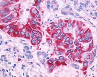
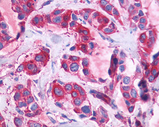
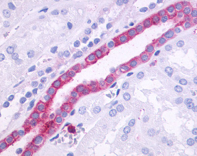
Application
| IHC-P |
|---|---|
| Primary Accession | Q5T601 |
| Reactivity | Human |
| Host | Rabbit |
| Clonality | Polyclonal |
| Calculated MW | 101kDa |
| Dilution | IHC-P (10-20 µg/ml) |
| Gene ID | 266977 |
|---|---|
| Other Names | Probable G-protein coupled receptor 110, G-protein coupled receptor KPG_012, G-protein coupled receptor PGR19, GPR110, PGR19 |
| Target/Specificity | Human GPR110. BLAST analysis of the peptide immunogen showed no homology with other human proteins. |
| Reconstitution & Storage | Long term: -70°C; Short term: +4°C |
| Precautions | ADGRF1 Antibody (N-Terminus) is for research use only and not for use in diagnostic or therapeutic procedures. |
| Name | ADGRF1 {ECO:0000303|PubMed:35418679, ECO:0000312|HGNC:HGNC:18990} |
|---|---|
| Function | Adhesion G-protein coupled receptor (aGPCR) for N- docosahexaenoylethanolamine (synaptamide), an omega-3 fatty acid lipid highly enriched in the brain (PubMed:27759003, PubMed:32144388). Ligand binding causes a conformation change that triggers signaling via guanine nucleotide-binding proteins (G proteins) and modulates the activity of downstream effectors, such as adenylate cyclase (PubMed:35418679, PubMed:36127364, PubMed:37120430). ADGRF1 is coupled to G(s) G proteins and mediates activation of adenylate cyclase activity (PubMed:35418679). Also able to couple to G(q), G(i) and G(12)/G(13) G proteins; additional evidence is however required to confirm this result in vivo (PubMed:36127364, PubMed:37120430). Involved in the development of neurons and cognitive function (By similarity). In liver, involved in fat accumulation (By similarity). |
| Cellular Location | Cell membrane; Multi-pass membrane protein |
| Tissue Location | Mainly expressed in the kidney. Up-regulated in lung adenocarcinomas and prostate cancers |
| Volume | 50 µl |

Thousands of laboratories across the world have published research that depended on the performance of antibodies from Abcepta to advance their research. Check out links to articles that cite our products in major peer-reviewed journals, organized by research category.
info@abcepta.com, and receive a free "I Love Antibodies" mug.

Provided below are standard protocols that you may find useful for product applications.
Background
Orphan receptor.
References
Suwa M.,et al.Submitted (JUL-2001) to the EMBL/GenBank/DDBJ databases.
Okazaki H.,et al.Submitted (JUN-2000) to the EMBL/GenBank/DDBJ databases.
Ota T.,et al.Nat. Genet. 36:40-45(2004).
Mungall A.J.,et al.Nature 425:805-811(2003).
Mural R.J.,et al.Submitted (JUL-2005) to the EMBL/GenBank/DDBJ databases.
If you have used an Abcepta product and would like to share how it has performed, please click on the "Submit Review" button and provide the requested information. Our staff will examine and post your review and contact you if needed.
If you have any additional inquiries please email technical services at tech@abcepta.com.
Ordering Information
Other Products
Shipping Information
