


 Foundational characteristics of cancer include proliferation, angiogenesis, migration, evasion of apoptosis, and cellular immortality. Find key markers for these cellular processes and antibodies to detect them.
 The SUMOplot™ Analysis Program predicts and scores sumoylation sites in your protein. SUMOylation is a post-translational modification involved in various cellular processes, such as nuclear-cytosolic transport, transcriptional regulation, apoptosis, protein stability, response to stress, and progression through the cell cycle.
 The Autophagy Receptor Motif Plotter predicts and scores autophagy receptor binding sites in your protein. Identifying proteins connected to this pathway is critical to understanding the role of autophagy in physiological as well as pathological processes such as development, differentiation, neurodegenerative diseases, stress, infection, and cancer.
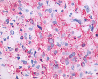
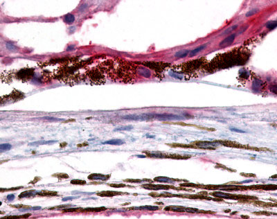
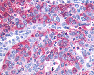
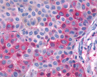
GPR143 Antibody (C-Terminus)
Rabbit Polyclonal Antibody
- SPECIFICATION
- CITATIONS
- PROTOCOLS
- BACKGROUND

Application
| IHC-P |
|---|---|
| Primary Accession | P51810 |
| Reactivity | Human |
| Host | Rabbit |
| Clonality | Polyclonal |
| Calculated MW | 44kDa |
| Dilution | IHC-P (6-14 µg/ml) |
| Gene ID | 4935 |
|---|---|
| Other Names | G-protein coupled receptor 143, Ocular albinism type 1 protein, GPR143, OA1 |
| Target/Specificity | Human GPR143. BLAST analysis of the peptide immunogen showed no homology with other human proteins. |
| Reconstitution & Storage | Long term: -70°C; Short term: +4°C |
| Precautions | GPR143 Antibody (C-Terminus) is for research use only and not for use in diagnostic or therapeutic procedures. |
| Name | GPR143 |
|---|---|
| Synonyms | OA1 |
| Function | Receptor for tyrosine, L-DOPA and dopamine. After binding to L-DOPA, stimulates Ca(2+) influx into the cytoplasm, increases secretion of the neurotrophic factor SERPINF1 and relocalizes beta arrestin at the plasma membrane; this ligand-dependent signaling occurs through a G(q)-mediated pathway in melanocytic cells. Its activity is mediated by G proteins which activate the phosphoinositide signaling pathway. Also plays a role as an intracellular G protein-coupled receptor involved in melanosome biogenesis, organization and transport. |
| Cellular Location | Melanosome membrane; Multi-pass membrane protein. Lysosome membrane; Multi-pass membrane protein. Apical cell membrane; Multi-pass membrane protein. Note=Distributed throughout the endo-melanosomal system but most of endogenous protein is localized in unpigmented stage II melanosomes. Its expression on the apical cell membrane is sensitive to tyrosine (PubMed:18828673). |
| Tissue Location | Expressed at high levels in the retina, including the retinal pigment epithelium (RPE), and in melanocytes. Weak expression is observed in brain and adrenal gland |
| Volume | 50 µl |

Thousands of laboratories across the world have published research that depended on the performance of antibodies from Abcepta to advance their research. Check out links to articles that cite our products in major peer-reviewed journals, organized by research category.
info@abcepta.com, and receive a free "I Love Antibodies" mug.

Provided below are standard protocols that you may find useful for product applications.
Background
Receptor for tyrosine, L-DOPA and dopamine. After binding to L-DOPA, stimulates Ca(2+) influx into the cytoplasm, increases secretion of the neurotrophic factor SERPINF1 and relocalizes beta arrestin at the plasma membrane; this ligand- dependent signaling occurs through a G(q)-mediated pathway in melanocytic cells. Its activity is mediated by G proteins which activate the phosphoinositide signaling pathway. Plays also a role as an intracellular G protein-coupled receptor involved in melanosome biogenesis, organization and transport.
References
Bassi M.T.,et al.Nat. Genet. 10:13-19(1995).
Ross M.T.,et al.Nature 434:325-337(2005).
Mural R.J.,et al.Submitted (JUL-2005) to the EMBL/GenBank/DDBJ databases.
Oetting W.S.,et al.Hum. Mutat. 13:99-115(1999).
Schiaffino M.V.,et al.Nat. Genet. 23:108-112(1999).
If you have used an Abcepta product and would like to share how it has performed, please click on the "Submit Review" button and provide the requested information. Our staff will examine and post your review and contact you if needed.
If you have any additional inquiries please email technical services at tech@abcepta.com.
Ordering Information
Shipping Information
