


 Foundational characteristics of cancer include proliferation, angiogenesis, migration, evasion of apoptosis, and cellular immortality. Find key markers for these cellular processes and antibodies to detect them.
 The SUMOplot™ Analysis Program predicts and scores sumoylation sites in your protein. SUMOylation is a post-translational modification involved in various cellular processes, such as nuclear-cytosolic transport, transcriptional regulation, apoptosis, protein stability, response to stress, and progression through the cell cycle.
 The Autophagy Receptor Motif Plotter predicts and scores autophagy receptor binding sites in your protein. Identifying proteins connected to this pathway is critical to understanding the role of autophagy in physiological as well as pathological processes such as development, differentiation, neurodegenerative diseases, stress, infection, and cancer.
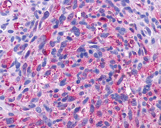
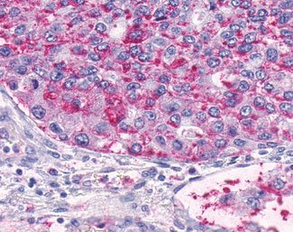
ADGRA2 / GPR124 Antibody (N-Terminus)
Rabbit Polyclonal Antibody
- SPECIFICATION
- CITATIONS
- PROTOCOLS
- BACKGROUND

Application
| IHC-P |
|---|---|
| Primary Accession | Q96PE1 |
| Reactivity | Human |
| Host | Rabbit |
| Clonality | Polyclonal |
| Calculated MW | 143kDa |
| Dilution | IHC-P (2-5 µg/ml) |
| Gene ID | 25960 |
|---|---|
| Other Names | G-protein coupled receptor 124, Tumor endothelial marker 5, GPR124, KIAA1531, TEM5 |
| Target/Specificity | Human GPR124. BLAST analysis of the peptide immunogen showed no homology with other human proteins. |
| Reconstitution & Storage | Long term: -70°C; Short term: +4°C |
| Precautions | ADGRA2 / GPR124 Antibody (N-Terminus) is for research use only and not for use in diagnostic or therapeutic procedures. |
| Name | ADGRA2 (HGNC:17849) |
|---|---|
| Function | Endothelial receptor which functions together with RECK to enable brain endothelial cells to selectively respond to Wnt7 signals (WNT7A or WNT7B) (PubMed:28289266, PubMed:30026314). Plays a key role in Wnt7-specific responses, such as endothelial cell sprouting and migration in the forebrain and neural tube, and establishment of the blood-brain barrier (By similarity). Acts as a Wnt7-specific coactivator of canonical Wnt signaling: required to deliver RECK-bound Wnt7 to frizzled by assembling a higher-order RECK-ADGRA2-Fzd-LRP5-LRP6 complex (PubMed:30026314). ADGRA2-tethering function does not rely on its G-protein coupled receptor (GPCR) structure but instead on its combined capacity to interact with RECK extracellularly and recruit the Dishevelled scaffolding protein intracellularly (PubMed:30026314). Binds to the glycosaminoglycans heparin, heparin sulfate, chondroitin sulfate and dermatan sulfate (PubMed:16982628). |
| Cellular Location | Cell membrane; Multi-pass membrane protein. Cell projection, filopodium. Note=Enriched at lateral cell borders and also at sites of cell-ECM (extracellular matrix) contact |
| Tissue Location | Expressed in endothelial cells (at protein level) (PubMed:15021905, PubMed:16982628). Abundantly expressed in heart, placenta, ovary, small intestine, and colon (PubMed:15021905) |
| Volume | 50 µl |

Thousands of laboratories across the world have published research that depended on the performance of antibodies from Abcepta to advance their research. Check out links to articles that cite our products in major peer-reviewed journals, organized by research category.
info@abcepta.com, and receive a free "I Love Antibodies" mug.

Provided below are standard protocols that you may find useful for product applications.
Background
Endothelial receptor that acts as an essential regulator of CNS angiogenesis. Required for normal endothelial cell sprouting and migration in the forebrain and neural tube (By similarity).
References
Carson-Walter E.B.,et al.Cancer Res. 61:6649-6655(2001).
Nagase T.,et al.DNA Res. 7:143-150(2000).
Nakajima D.,et al.DNA Res. 9:99-106(2002).
Ota T.,et al.Nat. Genet. 36:40-45(2004).
Nusbaum C.,et al.Nature 439:331-335(2006).
If you have used an Abcepta product and would like to share how it has performed, please click on the "Submit Review" button and provide the requested information. Our staff will examine and post your review and contact you if needed.
If you have any additional inquiries please email technical services at tech@abcepta.com.
Ordering Information
Other Products
Shipping Information
