


 Foundational characteristics of cancer include proliferation, angiogenesis, migration, evasion of apoptosis, and cellular immortality. Find key markers for these cellular processes and antibodies to detect them.
 The SUMOplot™ Analysis Program predicts and scores sumoylation sites in your protein. SUMOylation is a post-translational modification involved in various cellular processes, such as nuclear-cytosolic transport, transcriptional regulation, apoptosis, protein stability, response to stress, and progression through the cell cycle.
 The Autophagy Receptor Motif Plotter predicts and scores autophagy receptor binding sites in your protein. Identifying proteins connected to this pathway is critical to understanding the role of autophagy in physiological as well as pathological processes such as development, differentiation, neurodegenerative diseases, stress, infection, and cancer.
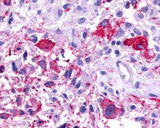
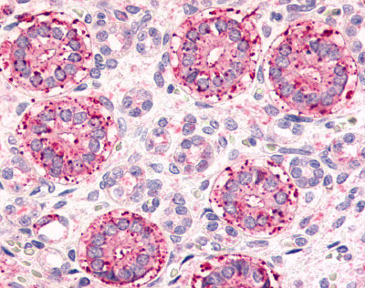
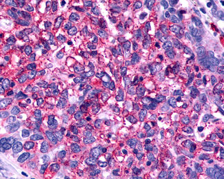
FSD2 / Frizzled 2 Antibody (N-Terminus)
Rabbit Polyclonal Antibody
- SPECIFICATION
- CITATIONS
- PROTOCOLS
- BACKGROUND

Application
| IHC-P |
|---|---|
| Primary Accession | Q14332 |
| Reactivity | Human, Mouse, Rabbit, Hamster, Monkey, Pig, Horse, Bovine, Dog |
| Host | Rabbit |
| Clonality | Polyclonal |
| Calculated MW | 64kDa |
| Dilution | IHC-P (8 µg/ml) |
| Gene ID | 2535 |
|---|---|
| Other Names | Frizzled-2, Fz-2, hFz2, FzE2, FZD2 |
| Target/Specificity | Human Frizzled 2. BLAST analysis of the peptide immunogen showed no homology with other human proteins, except FZD7 (60%), DMAP1 (40%). |
| Reconstitution & Storage | Long term: -70°C; Short term: +4°C |
| Precautions | FSD2 / Frizzled 2 Antibody (N-Terminus) is for research use only and not for use in diagnostic or therapeutic procedures. |
| Name | FZD2 |
|---|---|
| Function | Receptor for Wnt proteins. Most of frizzled receptors are coupled to the beta-catenin canonical signaling pathway, which leads to the activation of disheveled proteins, inhibition of GSK-3 kinase, nuclear accumulation of beta-catenin and activation of Wnt target genes (PubMed:25759469). A second signaling pathway involving PKC and calcium fluxes has been seen for some family members, but it is not yet clear if it represents a distinct pathway or if it can be integrated in the canonical pathway, as PKC seems to be required for Wnt-mediated inactivation of GSK-3 kinase. Both pathways seem to involve interactions with G-proteins. May be involved in transduction and intercellular transmission of polarity information during tissue morphogenesis and/or in differentiated tissues. |
| Cellular Location | Membrane; Multi-pass membrane protein. Cell membrane; Multi-pass membrane protein |
| Tissue Location | Widely expressed. In the adult, mainly found in heart, placenta, skeletal muscle, lung, kidney, pancreas, prostate, testis, ovary and colon. In the fetus, expressed in brain, lung and kidney. Low levels in fetal liver |
| Volume | 50 µl |

Thousands of laboratories across the world have published research that depended on the performance of antibodies from Abcepta to advance their research. Check out links to articles that cite our products in major peer-reviewed journals, organized by research category.
info@abcepta.com, and receive a free "I Love Antibodies" mug.

Provided below are standard protocols that you may find useful for product applications.
Background
Receptor for Wnt proteins. Most of frizzled receptors are coupled to the beta-catenin canonical signaling pathway, which leads to the activation of disheveled proteins, inhibition of GSK- 3 kinase, nuclear accumulation of beta-catenin and activation of Wnt target genes. A second signaling pathway involving PKC and calcium fluxes has been seen for some family members, but it is not yet clear if it represents a distinct pathway or if it can be integrated in the canonical pathway, as PKC seems to be required for Wnt-mediated inactivation of GSK-3 kinase. Both pathways seem to involve interactions with G-proteins. May be involved in transduction and intercellular transmission of polarity information during tissue morphogenesis and/or in differentiated tissues.
References
Zhao Z.,et al.Genomics 27:370-373(1995).
Sagara N.,et al.Biochem. Biophys. Res. Commun. 252:117-122(1998).
Tanaka S.,et al.Proc. Natl. Acad. Sci. U.S.A. 95:10164-10169(1998).
If you have used an Abcepta product and would like to share how it has performed, please click on the "Submit Review" button and provide the requested information. Our staff will examine and post your review and contact you if needed.
If you have any additional inquiries please email technical services at tech@abcepta.com.
Ordering Information
Other Products
Shipping Information
