


 Foundational characteristics of cancer include proliferation, angiogenesis, migration, evasion of apoptosis, and cellular immortality. Find key markers for these cellular processes and antibodies to detect them.
 The SUMOplot™ Analysis Program predicts and scores sumoylation sites in your protein. SUMOylation is a post-translational modification involved in various cellular processes, such as nuclear-cytosolic transport, transcriptional regulation, apoptosis, protein stability, response to stress, and progression through the cell cycle.
 The Autophagy Receptor Motif Plotter predicts and scores autophagy receptor binding sites in your protein. Identifying proteins connected to this pathway is critical to understanding the role of autophagy in physiological as well as pathological processes such as development, differentiation, neurodegenerative diseases, stress, infection, and cancer.
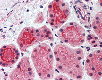
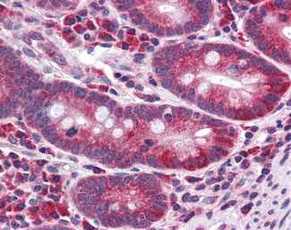
CDHR5 / MUCDHL Antibody (Cytoplasmic Domain)
Rabbit Polyclonal Antibody
- SPECIFICATION
- CITATIONS
- PROTOCOLS
- BACKGROUND

Application
| IHC-P |
|---|---|
| Primary Accession | Q9HBB8 |
| Reactivity | Human |
| Host | Rabbit |
| Clonality | Polyclonal |
| Calculated MW | 88kDa |
| Dilution | IHC-P (5-7.5 µg/ml) |
| Gene ID | 53841 |
|---|---|
| Other Names | Cadherin-related family member 5, Mu-protocadherin, Mucin and cadherin-like protein, CDHR5, MUCDHL, MUPCDH |
| Target/Specificity | Human CDHR5 / MUPCDH. BLAST analysis of the peptide immunogen showed no homology with other human proteins. |
| Reconstitution & Storage | Long term: -70°C; Short term: +4°C |
| Precautions | CDHR5 / MUCDHL Antibody (Cytoplasmic Domain) is for research use only and not for use in diagnostic or therapeutic procedures. |
| Name | CDHR5 (HGNC:7521) |
|---|---|
| Function | Intermicrovillar adhesion molecule that forms, via its extracellular domain, calcium-dependent heterophilic complexes with CDHR2 on adjacent microvilli. Thereby, controls the packing of microvilli at the apical membrane of epithelial cells. Through its cytoplasmic domain, interacts with microvillus cytoplasmic proteins to form the intermicrovillar adhesion complex/IMAC. This complex plays a central role in microvilli and epithelial brush border differentiation. |
| Cellular Location | Apical cell membrane; Single-pass type I membrane protein. Cell projection, microvillus membrane; Single-pass type I membrane protein |
| Tissue Location | Highest expression in kidney, liver, colon and small intestine. In kidney, expressed apically along brush border of proximal convoluted tubule but not in cortical collecting ducts Isoform 1 is expressed primarily in adult small intestine and colon Isoform 2 is highly expressed in fetal liver (PubMed:12167596) Expressed in duodenum with higher expression in enterocytes along the villus axis and lower expression in crypts (at protein level) (PubMed:24725409). |

Thousands of laboratories across the world have published research that depended on the performance of antibodies from Abcepta to advance their research. Check out links to articles that cite our products in major peer-reviewed journals, organized by research category.
info@abcepta.com, and receive a free "I Love Antibodies" mug.

Provided below are standard protocols that you may find useful for product applications.
Background
Acts as a calcium-dependent cell adhesion protein.
References
Paris M.J.,et al.Genomics 69:196-202(2000).
Soleiman A.,et al.Submitted (AUG-2000) to the EMBL/GenBank/DDBJ databases.
Clark H.F.,et al.Genome Res. 13:2265-2270(2003).
Ota T.,et al.Nat. Genet. 36:40-45(2004).
Taylor T.D.,et al.Nature 440:497-500(2006).
If you have used an Abcepta product and would like to share how it has performed, please click on the "Submit Review" button and provide the requested information. Our staff will examine and post your review and contact you if needed.
If you have any additional inquiries please email technical services at tech@abcepta.com.
Ordering Information
Other Products
Shipping Information
