


 Foundational characteristics of cancer include proliferation, angiogenesis, migration, evasion of apoptosis, and cellular immortality. Find key markers for these cellular processes and antibodies to detect them.
 The SUMOplot™ Analysis Program predicts and scores sumoylation sites in your protein. SUMOylation is a post-translational modification involved in various cellular processes, such as nuclear-cytosolic transport, transcriptional regulation, apoptosis, protein stability, response to stress, and progression through the cell cycle.
 The Autophagy Receptor Motif Plotter predicts and scores autophagy receptor binding sites in your protein. Identifying proteins connected to this pathway is critical to understanding the role of autophagy in physiological as well as pathological processes such as development, differentiation, neurodegenerative diseases, stress, infection, and cancer.
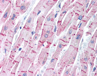
STEAP4 Antibody (Internal)
Rabbit Polyclonal Antibody
- SPECIFICATION
- CITATIONS
- PROTOCOLS
- BACKGROUND

Application
| WB, IHC-P |
|---|---|
| Primary Accession | Q687X5 |
| Reactivity | Human, Mouse, Rat |
| Host | Rabbit |
| Clonality | Polyclonal |
| Calculated MW | 52kDa |
| Dilution | IHC-P (5 µg/ml), WB (2 µg/ml), |
| Gene ID | 79689 |
|---|---|
| Other Names | Metalloreductase STEAP4, 1.16.1.-, Six-transmembrane epithelial antigen of prostate 4, SixTransMembrane protein of prostate 2, Tumor necrosis factor, alpha-induced protein 9, STEAP4, STAMP2, TNFAIP9 |
| Target/Specificity | a 14 amino acid peptide from near the center of human STEAP4 |
| Reconstitution & Storage | Short term 4°C, long term aliquot and store at -20°C, avoid freeze thaw cycles. Store undiluted. |
| Precautions | STEAP4 Antibody (Internal) is for research use only and not for use in diagnostic or therapeutic procedures. |
| Name | STEAP4 |
|---|---|
| Synonyms | STAMP2 {ECO:0000303|PubMed:15897894}, TN |
| Function | Integral membrane protein that functions as a NADPH-dependent ferric-chelate reductase, using NADPH from one side of the membrane to reduce a Fe(3+) chelate that is bound on the other side of the membrane. Mediates sequential transmembrane electron transfer from NADPH to FAD and onto heme, and finally to the Fe(3+) chelate (PubMed:30337524). Can also reduce Cu(2+) to Cu(1+) (By similarity). Plays a role in systemic metabolic homeostasis, integrating inflammatory and metabolic responses (By similarity). Associated with obesity and insulin-resistance (PubMed:18381574, PubMed:18430367). Involved in inflammatory arthritis, through the regulation of inflammatory cytokines (PubMed:19660107). Inhibits anchorage- independent cell proliferation (PubMed:19787193). |
| Cellular Location | Cell membrane; Multi-pass membrane protein. Golgi apparatus membrane; Multi-pass membrane protein. Early endosome membrane; Multi-pass membrane protein |
| Tissue Location | Ubiquitous. Highly expressed in adipose tissue. Expressed in placenta, lung, heart and prostate. Detected at lower levels in liver, skeletal muscle, pancreas, testis and small intestine Highly expressed in joints of patients with rheumatoid arthritis and localized with CD68 cells, a marker for macrophages |

Thousands of laboratories across the world have published research that depended on the performance of antibodies from Abcepta to advance their research. Check out links to articles that cite our products in major peer-reviewed journals, organized by research category.
info@abcepta.com, and receive a free "I Love Antibodies" mug.

Provided below are standard protocols that you may find useful for product applications.
Background
Metalloreductase that has the ability to reduce both Fe(3+) to Fe(2+) and Cu(2+) to Cu(1+). Uses NAD(+) as acceptor. Plays a role in systemic metabolic homeostasis, integrating inflammatory and metabolic responses (By similarity). Associated with obesity and insulin-resistance. Involved in inflammatory arthritis, through the regulation of inflammatory cytokines. Inhibits anchorage-independent cell proliferation.
References
Korkmaz C.G.,et al.Oncogene 24:4934-4945(2005).
Ota T.,et al.Nat. Genet. 36:40-45(2004).
Bechtel S.,et al.BMC Genomics 8:399-399(2007).
Ohgami R.S.,et al.Nat. Genet. 37:1264-1269(2005).
Zhang C.M.,et al.Acta Pharmacol. Sin. 29:587-592(2008).
If you have used an Abcepta product and would like to share how it has performed, please click on the "Submit Review" button and provide the requested information. Our staff will examine and post your review and contact you if needed.
If you have any additional inquiries please email technical services at tech@abcepta.com.
Ordering Information
Other Products
Shipping Information
