


 Foundational characteristics of cancer include proliferation, angiogenesis, migration, evasion of apoptosis, and cellular immortality. Find key markers for these cellular processes and antibodies to detect them.
 The SUMOplot™ Analysis Program predicts and scores sumoylation sites in your protein. SUMOylation is a post-translational modification involved in various cellular processes, such as nuclear-cytosolic transport, transcriptional regulation, apoptosis, protein stability, response to stress, and progression through the cell cycle.
 The Autophagy Receptor Motif Plotter predicts and scores autophagy receptor binding sites in your protein. Identifying proteins connected to this pathway is critical to understanding the role of autophagy in physiological as well as pathological processes such as development, differentiation, neurodegenerative diseases, stress, infection, and cancer.
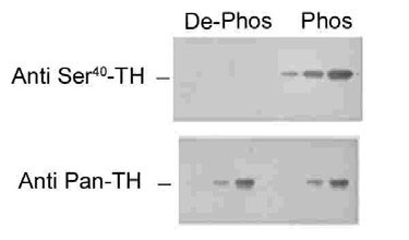
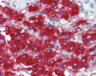
TH / Tyrosine Hydroxylase Antibody (phospho-Ser40)
Rabbit Polyclonal Antibody
- SPECIFICATION
- CITATIONS
- PROTOCOLS
- BACKGROUND

Application
| WB, IHC-P, IF, IHC-F |
|---|---|
| Primary Accession | P07101 |
| Reactivity | Human, Rat |
| Host | Rabbit |
| Clonality | Polyclonal |
| Calculated MW | 59kDa |
| Dilution | IF (1:1000), IHC-Fr (1:1000), IHC-P (5 µg/ml), WB (1:1000) , |
| Gene ID | 7054 |
|---|---|
| Other Names | Tyrosine 3-monooxygenase, 1.14.16.2, Tyrosine 3-hydroxylase, TH, TH, TYH |
| Target/Specificity | Specific for the ~60k tyrosine hydroxylase protein phosphorylated at Ser40. Some higher molecular weight bands may be detected by the antibody depending upon the brain region being studied, protein loads and the detection methods used. The antibody h ... |
| Reconstitution & Storage | Long term: -80°C; Short term: -20°C |
| Precautions | TH / Tyrosine Hydroxylase Antibody (phospho-Ser40) is for research use only and not for use in diagnostic or therapeutic procedures. |
| Name | TH (HGNC:11782) |
|---|---|
| Synonyms | TYH |
| Function | Catalyzes the conversion of L-tyrosine to L- dihydroxyphenylalanine (L-Dopa), the rate-limiting step in the biosynthesis of catecholamines, dopamine, noradrenaline, and adrenaline. Uses tetrahydrobiopterin and molecular oxygen to convert tyrosine to L-Dopa (PubMed:15287903, PubMed:1680128, PubMed:17391063, PubMed:24753243, PubMed:34922205, PubMed:8528210, Ref.18). In addition to tyrosine, is able to catalyze the hydroxylation of phenylalanine and tryptophan with lower specificity (By similarity). Positively regulates the regression of retinal hyaloid vessels during postnatal development (By similarity). |
| Cellular Location | Cytoplasm, perinuclear region {ECO:0000250|UniProtKB:P24529}. Nucleus {ECO:0000250|UniProtKB:P04177} Cell projection, axon {ECO:0000250|UniProtKB:P24529}. Cytoplasm {ECO:0000250|UniProtKB:P04177}. Cytoplasmic vesicle, secretory vesicle, synaptic vesicle {ECO:0000250|UniProtKB:P04177}. Note=When phosphorylated at Ser-19 shows a nuclear distribution and when phosphorylated at Ser-31 as well at Ser-40 shows a cytosolic distribution (By similarity). Expressed in dopaminergic axons and axon terminals. {ECO:0000250|UniProtKB:P04177} |
| Tissue Location | Mainly expressed in the brain and adrenal glands. |

Thousands of laboratories across the world have published research that depended on the performance of antibodies from Abcepta to advance their research. Check out links to articles that cite our products in major peer-reviewed journals, organized by research category.
info@abcepta.com, and receive a free "I Love Antibodies" mug.

Provided below are standard protocols that you may find useful for product applications.
Background
Plays an important role in the physiology of adrenergic neurons.
References
Kaneda N.,et al.Biochem. Biophys. Res. Commun. 146:971-975(1987).
Grima B.,et al.Nature 326:707-711(1987).
Kobayashi K.,et al.Nucleic Acids Res. 15:6733-6733(1987).
Kobayashi K.,et al.J. Biochem. 103:907-912(1988).
Roma J.,et al.Biol. Chem. 388:419-426(2007).
If you have used an Abcepta product and would like to share how it has performed, please click on the "Submit Review" button and provide the requested information. Our staff will examine and post your review and contact you if needed.
If you have any additional inquiries please email technical services at tech@abcepta.com.
Ordering Information
Other Products
Shipping Information
