


 Foundational characteristics of cancer include proliferation, angiogenesis, migration, evasion of apoptosis, and cellular immortality. Find key markers for these cellular processes and antibodies to detect them.
 The SUMOplot™ Analysis Program predicts and scores sumoylation sites in your protein. SUMOylation is a post-translational modification involved in various cellular processes, such as nuclear-cytosolic transport, transcriptional regulation, apoptosis, protein stability, response to stress, and progression through the cell cycle.
 The Autophagy Receptor Motif Plotter predicts and scores autophagy receptor binding sites in your protein. Identifying proteins connected to this pathway is critical to understanding the role of autophagy in physiological as well as pathological processes such as development, differentiation, neurodegenerative diseases, stress, infection, and cancer.
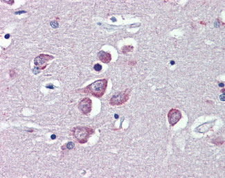
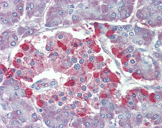
TRPM2 Antibody (Internal)
Goat Polyclonal Antibody
- SPECIFICATION
- CITATIONS
- PROTOCOLS
- BACKGROUND

Application
| IHC-P |
|---|---|
| Primary Accession | O94759 |
| Reactivity | Human, Monkey |
| Host | Goat |
| Clonality | Polyclonal |
| Calculated MW | 171kDa |
| Dilution | IHC-P (3.75 µg/ml) |
| Gene ID | 7226 |
|---|---|
| Other Names | Transient receptor potential cation channel subfamily M member 2, 3.6.1.13, Estrogen-responsive element-associated gene 1 protein, Long transient receptor potential channel 2, LTrpC-2, LTrpC2, Transient receptor potential channel 7, TrpC7, TRPM2, EREG1, KNP3, LTRPC2, TRPC7 |
| Target/Specificity | Human TRPM2. |
| Reconstitution & Storage | Store at -20°C. Minimize freezing and thawing. |
| Precautions | TRPM2 Antibody (Internal) is for research use only and not for use in diagnostic or therapeutic procedures. |
| Name | TRPM2 |
|---|---|
| Function | [Isoform 1]: Nonselective, voltage-independent cation channel that mediates Na(+) and Ca(2+) influx, leading to increased cytoplasmic Ca(2+) levels (PubMed:11960981, PubMed:12594222, PubMed:11385575, PubMed:11509734, PubMed:11804595, PubMed:15561722, PubMed:16601673, PubMed:19171771, PubMed:20660597, PubMed:25620041, PubMed:27383051, PubMed:27068538, PubMed:28775320, PubMed:29745897, PubMed:30467180). Functions as a ligand-gated ion channel (PubMed:19171771, PubMed:25620041, PubMed:28775320, PubMed:30467180). Binding of ADP- ribose to the cytoplasmic Nudix domain causes a conformation change; the channel is primed but still requires Ca(2+) binding to trigger channel opening (PubMed:19171771, PubMed:25620041, PubMed:28775320, PubMed:29745897, PubMed:30467180). Extracellular calcium passes through the channel and increases channel activity (PubMed:19171771). Contributes to Ca(2+) release from intracellular stores in response to ADP-ribose (PubMed:19454650). Plays a role in numerous processes that involve signaling via intracellular Ca(2+) levels (Probable). Besides, mediates the release of lysosomal Zn(2+) stores in response to reactive oxygen species, leading to increased cytosolic Zn(2+) levels (PubMed:25562606, PubMed:27068538). Activated by moderate heat (35 to 40 degrees Celsius) (PubMed:16601673). Activated by intracellular ADP- ribose, beta-NAD (NAD(+)) and similar compounds, and by oxidative stress caused by reactive oxygen or nitrogen species (PubMed:11960981, PubMed:11385575, PubMed:11509734, PubMed:11804595, PubMed:15561722, PubMed:16601673, PubMed:19171771, PubMed:25620041, PubMed:27383051, PubMed:27068538, PubMed:30467180). The precise physiological activators are under debate; the true, physiological activators may be ADP-ribose and ADP-ribose-2'-phosphate (PubMed:20650899, PubMed:25918360). Activation by ADP-ribose and beta-NAD is strongly increased by moderate heat (35 to 40 degrees Celsius) (PubMed:16601673). Likewise, reactive oxygen species lower the threshold for activation by moderate heat (37 degrees Celsius) (PubMed:22493272). Plays a role in mediating behavorial and physiological responses to moderate heat and thereby contributes to body temperature homeostasis. Plays a role in insulin secretion, a process that requires increased cytoplasmic Ca(2+) levels (By similarity). Required for normal IFNG and cytokine secretion and normal innate immune immunity in response to bacterial infection. Required for normal phagocytosis and cytokine release by macrophages exposed to zymosan (in vitro). Plays a role in dendritic cell differentiation and maturation, and in dendritic cell chemotaxis via its role in regulating cytoplasmic Ca(2+) levels (By similarity). Plays a role in the regulation of the reorganization of the actin cytoskeleton and filopodia formation in response to reactive oxygen species via its role in increasing cytoplasmic Ca(2+) and Zn(2+) levels (PubMed:27068538). Confers susceptibility to cell death following oxidative stress (PubMed:12594222, PubMed:25562606). |
| Cellular Location | Cell membrane; Multi-pass membrane protein. Perikaryon {ECO:0000250|UniProtKB:E9PTA2}. Cell projection {ECO:0000250|UniProtKB:E9PTA2}. Cytoplasmic vesicle {ECO:0000250|UniProtKB:E9PTA2}. Lysosome Note=Detected at the cell membrane and in intracellular vesicles in cortical neurons. Detected on neuronal cell bodies and neurites (By similarity). Detected on the cell membrane in polymorphonuclear neutrophils. Detected on cytoplasmic vesicles and lysosomes in immature bone marrow dendritic cells (By similarity) {ECO:0000250|UniProtKB:E9PTA2, ECO:0000250|UniProtKB:Q91YD4} [Isoform 2]: Cell membrane; Multi-pass membrane protein |
| Tissue Location | Highly expressed in brain and peripheral blood cells, such as neutrophils. Also detected in bone marrow, spleen, heart, liver and lung. Isoform 2 is found in neutrophil granulocytes |
| Volume | 50 µl |

Thousands of laboratories across the world have published research that depended on the performance of antibodies from Abcepta to advance their research. Check out links to articles that cite our products in major peer-reviewed journals, organized by research category.
info@abcepta.com, and receive a free "I Love Antibodies" mug.

Provided below are standard protocols that you may find useful for product applications.
Background
Nonselective, voltage-independent cation channel mediating sodium and calcium ion influx in response to oxidative stress. Extracellular calcium passes through the channel and acts from the intracellular side as a positive regulator in channel activation. Activated by ADP-ribose, nicotinamide adenine dinucleotide (NAD(+)), reactive nitrogen species and arachidonic acid. Inactivated by intracellular ATP. Confers susceptibility to cell death following oxidative stress. Isoform 2 does not seem to be regulated by ADPR. Has ADP-ribose pyrophosphatase activity.
References
Nagamine K.,et al.Genomics 54:124-131(1998).
Wehage E.,et al.J. Biol. Chem. 277:23150-23156(2002).
Zhang W.,et al.J. Biol. Chem. 278:16222-16229(2003).
Uemura T.,et al.Biochem. Biophys. Res. Commun. 328:1232-1243(2005).
Hayes P.D.,et al.Submitted (DEC-2005) to the EMBL/GenBank/DDBJ databases.
If you have used an Abcepta product and would like to share how it has performed, please click on the "Submit Review" button and provide the requested information. Our staff will examine and post your review and contact you if needed.
If you have any additional inquiries please email technical services at tech@abcepta.com.
Ordering Information
Shipping Information
