


 Foundational characteristics of cancer include proliferation, angiogenesis, migration, evasion of apoptosis, and cellular immortality. Find key markers for these cellular processes and antibodies to detect them.
 The SUMOplot™ Analysis Program predicts and scores sumoylation sites in your protein. SUMOylation is a post-translational modification involved in various cellular processes, such as nuclear-cytosolic transport, transcriptional regulation, apoptosis, protein stability, response to stress, and progression through the cell cycle.
 The Autophagy Receptor Motif Plotter predicts and scores autophagy receptor binding sites in your protein. Identifying proteins connected to this pathway is critical to understanding the role of autophagy in physiological as well as pathological processes such as development, differentiation, neurodegenerative diseases, stress, infection, and cancer.
CDKN2A / p16INK4a Antibody (clone 1E12E10)
Mouse Monoclonal Antibody
- SPECIFICATION
- CITATIONS
- PROTOCOLS
- BACKGROUND

Application
| WB, IHC-P, E |
|---|---|
| Primary Accession | P42771 |
| Reactivity | Human, Mouse, Rat |
| Host | Mouse |
| Clonality | Monoclonal |
| Isotype | IgG1 Monoclonal [1E12E10] |
| Clone Names | 1E12E10 |
| Calculated MW | 17kDa |
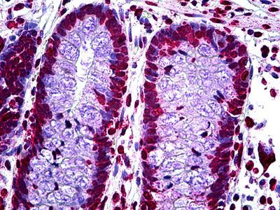
| Dilution | ELISA (1:10000), IHC-P (5 µg/ml), WB (1:500-1:2000) |
| Gene ID | 1029 |
|---|---|
| Other Names | Cyclin-dependent kinase inhibitor 2A, isoforms 1/2/3, Cyclin-dependent kinase 4 inhibitor A, CDK4I, Multiple tumor suppressor 1, MTS-1, p16-INK4a, p16-INK4, p16INK4A, CDKN2A, CDKN2, MTS1 |
| Target/Specificity | Targets P16. Shows reactivity with Human, Mouse, and Rat samples. |
| Format | PBS with 0.1% sodium azide and 1% BSA. |
| Reconstitution & Storage | Long term: -20°C; Short term: +4°C. Avoid repeat freeze-thaw cycles. |
| Precautions | CDKN2A / p16INK4a Antibody (clone 1E12E10) is for research use only and not for use in diagnostic or therapeutic procedures. |
| Name | CDKN2A (HGNC:1787) |
|---|---|
| Synonyms | CDKN2, MTS1 |
| Function | Acts as a negative regulator of the proliferation of normal cells by interacting strongly with CDK4 and CDK6. This inhibits their ability to interact with cyclins D and to phosphorylate the retinoblastoma protein. |
| Cellular Location | Cytoplasm. Nucleus |
| Tissue Location | Widely expressed but not detected in brain or skeletal muscle. Isoform 3 is pancreas-specific |
| Volume | Array |

Thousands of laboratories across the world have published research that depended on the performance of antibodies from Abcepta to advance their research. Check out links to articles that cite our products in major peer-reviewed journals, organized by research category.
info@abcepta.com, and receive a free "I Love Antibodies" mug.

Provided below are standard protocols that you may find useful for product applications.
Background
Acts as a negative regulator of the proliferation of normal cells by interacting strongly with CDK4 and CDK6. This inhibits their ability to interact with cyclins D and to phosphorylate the retinoblastoma protein.
References
Serrano M.,et al.Nature 366:704-707(1993).
Robertson K.D.,et al.Oncogene 18:3810-3820(1999).
Kitagawa Y.,et al.J. Biol. Chem. 277:46289-46297(2002).
Lin Y.C.,et al.Oncogene 26:7017-7027(2007).
Humphray S.J.,et al.Nature 429:369-374(2004).
If you have used an Abcepta product and would like to share how it has performed, please click on the "Submit Review" button and provide the requested information. Our staff will examine and post your review and contact you if needed.
If you have any additional inquiries please email technical services at tech@abcepta.com.
Ordering Information
Other Products
Shipping Information
