


 Foundational characteristics of cancer include proliferation, angiogenesis, migration, evasion of apoptosis, and cellular immortality. Find key markers for these cellular processes and antibodies to detect them.
 The SUMOplot™ Analysis Program predicts and scores sumoylation sites in your protein. SUMOylation is a post-translational modification involved in various cellular processes, such as nuclear-cytosolic transport, transcriptional regulation, apoptosis, protein stability, response to stress, and progression through the cell cycle.
 The Autophagy Receptor Motif Plotter predicts and scores autophagy receptor binding sites in your protein. Identifying proteins connected to this pathway is critical to understanding the role of autophagy in physiological as well as pathological processes such as development, differentiation, neurodegenerative diseases, stress, infection, and cancer.
CLIC5 Antibody (clone 1E6)
Mouse Monoclonal Antibody
- SPECIFICATION
- CITATIONS
- PROTOCOLS
- BACKGROUND

Application
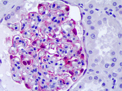
| WB, IHC-P, E |
|---|---|
| Primary Accession | Q9NZA1 |
| Reactivity | Human |
| Host | Mouse |
| Clonality | Monoclonal |
| Clone Names | 1E6 |
| Calculated MW | 47kDa |
| Dilution | IHC-P (10 µg/ml), WB (1:500-1:1000), |
| Gene ID | 53405 |
|---|---|
| Other Names | Chloride intracellular channel protein 5, CLIC5 |
| Target/Specificity | Human CLIC5 |
| Reconstitution & Storage | Short term 4°C, long term aliquot and store at -20°C, avoid freeze thaw cycles. |
| Precautions | CLIC5 Antibody (clone 1E6) is for research use only and not for use in diagnostic or therapeutic procedures. |
| Name | CLIC5 {ECO:0000303|PubMed:10793131, ECO:0000312|HGNC:HGNC:13517} |
|---|---|
| Function | In the soluble state, catalyzes glutaredoxin-like thiol disulfide exchange reactions with reduced glutathione as electron donor (By similarity). Can insert into membranes and form non-selective ion channels almost equally permeable to Na(+), K(+) and Cl(-) (PubMed:15184393, PubMed:18028448). Required for normal hearing (PubMed:24781754). It is necessary for the formation of stereocilia in the inner ear and normal development of the organ of Corti (By similarity). May play a role in the regulation of transepithelial ion absorption and secretion. Is required for the development and/or maintenance of the proper glomerular endothelial cell and podocyte architecture (PubMed:15184393, PubMed:18028448, PubMed:20335315). Plays a role in formation of the lens suture in the eye, which is important for normal optical properties of the lens (By similarity). |
| Cellular Location | [Isoform 1]: Cytoplasm, cytoskeleton. Cytoplasm, cell cortex. Membrane; Single-pass membrane protein. Apical cell membrane; Single-pass membrane protein. Cytoplasm {ECO:0000250|UniProtKB:O00299}. Mitochondrion {ECO:0000250|UniProtKB:Q9EPT8}. Cell projection, stereocilium. Note=Associates with the cortical actin cytoskeleton (PubMed:10793131, PubMed:15184393). Localizes to the apical region of cochlear hair cells, at the base of the actin-rich hair bundle (By similarity). Colocalizes with podocalyxin at the apical cell membrane in renal glomeruli (PubMed:20335315). May localize to the centrosome in lens epithelial cells (By similarity). Exists both as soluble cytoplasmic protein and as membrane protein with probably a single transmembrane domain (By similarity) {ECO:0000250|UniProtKB:O00299, ECO:0000250|UniProtKB:Q8BXK9, ECO:0000250|UniProtKB:Q9EPT8, ECO:0000269|PubMed:10793131, ECO:0000269|PubMed:15184393, ECO:0000269|PubMed:20335315} |
| Tissue Location | Widely expressed in both fetal and adult human tissues (PubMed:24781754). Isoform 1 is expressed in renal glomeruli endothelial cells and podocytes (at protein level) |

Thousands of laboratories across the world have published research that depended on the performance of antibodies from Abcepta to advance their research. Check out links to articles that cite our products in major peer-reviewed journals, organized by research category.
info@abcepta.com, and receive a free "I Love Antibodies" mug.

Provided below are standard protocols that you may find useful for product applications.
Background
Required for normal hearing (PubMed:24781754). It is necessary for the formation of stereocilia in the inner ear and normal development of the organ of Corti (By similarity). Can insert into membranes and form poorly selective ion channels that may also transport chloride ions. May play a role in the regulation of transepithelial ion absorption and secretion. Is required for the development and/or maintenance of the proper glomerular endothelial cell and podocyte architecture (PubMed:15184393, PubMed:18028448, PubMed:20335315).
References
Berryman M.,et al.Mol. Biol. Cell 11:1509-1521(2000).
Shanks R.A.,et al.J. Biol. Chem. 277:40973-40980(2002).
Ota T.,et al.Nat. Genet. 36:40-45(2004).
Otsuki T.,et al.DNA Res. 12:117-126(2005).
Mungall A.J.,et al.Nature 425:805-811(2003).
If you have used an Abcepta product and would like to share how it has performed, please click on the "Submit Review" button and provide the requested information. Our staff will examine and post your review and contact you if needed.
If you have any additional inquiries please email technical services at tech@abcepta.com.
Ordering Information
Other Products
Shipping Information
