


 Foundational characteristics of cancer include proliferation, angiogenesis, migration, evasion of apoptosis, and cellular immortality. Find key markers for these cellular processes and antibodies to detect them.
 The SUMOplot™ Analysis Program predicts and scores sumoylation sites in your protein. SUMOylation is a post-translational modification involved in various cellular processes, such as nuclear-cytosolic transport, transcriptional regulation, apoptosis, protein stability, response to stress, and progression through the cell cycle.
 The Autophagy Receptor Motif Plotter predicts and scores autophagy receptor binding sites in your protein. Identifying proteins connected to this pathway is critical to understanding the role of autophagy in physiological as well as pathological processes such as development, differentiation, neurodegenerative diseases, stress, infection, and cancer.
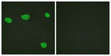
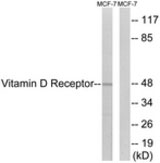
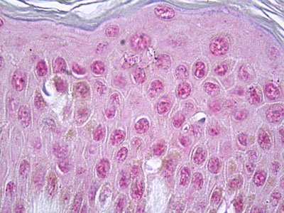
Vitamin D Receptor / VDR Antibody (aa181-230)
Rabbit Polyclonal Antibody
- SPECIFICATION
- CITATIONS
- PROTOCOLS
- BACKGROUND

Application
| WB, IHC-P, IF, E |
|---|---|
| Primary Accession | P11473 |
| Reactivity | Human |
| Host | Rabbit |
| Clonality | Polyclonal |
| Calculated MW | 48kDa |
| Dilution | ELISA (1:10000), IF (1:100-1:500), IHC-P (5 µg/ml), WB (1:500-1:1000) , |
| Gene ID | 7421 |
|---|---|
| Other Names | Vitamin D3 receptor, VDR, 1, 25-dihydroxyvitamin D3 receptor, Nuclear receptor subfamily 1 group I member 1, VDR, NR1I1 |
| Target/Specificity | Vitamin D Receptor (Ab-208) Antibody detects endogenous levels of total Vitamin D Receptor protein. |
| Reconstitution & Storage | Short term 4°C, long term aliquot and store at -20°C, avoid freeze thaw cycles. |
| Precautions | Vitamin D Receptor / VDR Antibody (aa181-230) is for research use only and not for use in diagnostic or therapeutic procedures. |
| Name | VDR (HGNC:12679) |
|---|---|
| Synonyms | NR1I1 |
| Function | Nuclear receptor for calcitriol, the active form of vitamin D3 which mediates the action of this vitamin on cells (PubMed:10678179, PubMed:15728261, PubMed:16913708, PubMed:28698609, PubMed:37478846). Enters the nucleus upon vitamin D3 binding where it forms heterodimers with the retinoid X receptor/RXR (PubMed:28698609). The VDR-RXR heterodimers bind to specific response elements on DNA and activate the transcription of vitamin D3-responsive target genes (PubMed:28698609). Plays a central role in calcium homeostasis (By similarity). Also functions as a receptor for the secondary bile acid lithocholic acid (LCA) and its metabolites (PubMed:12016314, PubMed:32354638). |
| Cellular Location | Nucleus {ECO:0000255|PROSITE-ProRule:PRU00407, ECO:0000269|PubMed:12145331, ECO:0000269|PubMed:16207705, ECO:0000269|PubMed:28698609}. Cytoplasm Note=Localizes mainly to the nucleus (PubMed:12145331, PubMed:28698609). Translocated into the nucleus via both ligand- dependent and ligand-independent pathways; ligand-independent nuclear translocation is mediated by IPO4 (PubMed:16207705) |
| Volume | 50 µl |

Thousands of laboratories across the world have published research that depended on the performance of antibodies from Abcepta to advance their research. Check out links to articles that cite our products in major peer-reviewed journals, organized by research category.
info@abcepta.com, and receive a free "I Love Antibodies" mug.

Provided below are standard protocols that you may find useful for product applications.
Background
Nuclear hormone receptor. Transcription factor that mediates the action of vitamin D3 by controlling the expression of hormone sensitive genes. Regulates transcription of hormone sensitive genes via its association with the WINAC complex, a chromatin-remodeling complex. Recruited to promoters via its interaction with the WINAC complex subunit BAZ1B/WSTF, which mediates the interaction with acetylated histones, an essential step for VDR-promoter association. Plays a central role in calcium homeostasis.
References
Baker A.R.,et al.Proc. Natl. Acad. Sci. U.S.A. 85:3294-3298(1988).
Goto H.,et al.Biochim. Biophys. Acta 1132:103-108(1992).
Rae J.L.,et al.Submitted (SEP-1997) to the EMBL/GenBank/DDBJ databases.
Miyamoto K.,et al.Mol. Endocrinol. 11:1165-1179(1997).
Ota T.,et al.Nat. Genet. 36:40-45(2004).
If you have used an Abcepta product and would like to share how it has performed, please click on the "Submit Review" button and provide the requested information. Our staff will examine and post your review and contact you if needed.
If you have any additional inquiries please email technical services at tech@abcepta.com.
Ordering Information
Other Products
Shipping Information
