


 Foundational characteristics of cancer include proliferation, angiogenesis, migration, evasion of apoptosis, and cellular immortality. Find key markers for these cellular processes and antibodies to detect them.
 The SUMOplot™ Analysis Program predicts and scores sumoylation sites in your protein. SUMOylation is a post-translational modification involved in various cellular processes, such as nuclear-cytosolic transport, transcriptional regulation, apoptosis, protein stability, response to stress, and progression through the cell cycle.
 The Autophagy Receptor Motif Plotter predicts and scores autophagy receptor binding sites in your protein. Identifying proteins connected to this pathway is critical to understanding the role of autophagy in physiological as well as pathological processes such as development, differentiation, neurodegenerative diseases, stress, infection, and cancer.
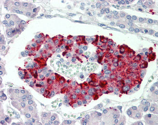
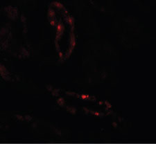
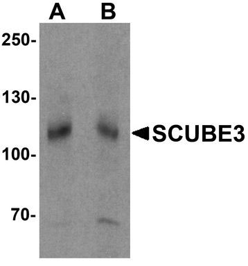
SCUBE3 Antibody (Internal)
Rabbit Polyclonal Antibody
- SPECIFICATION
- CITATIONS
- PROTOCOLS
- BACKGROUND

Application
| WB, IHC-P, IF |
|---|---|
| Primary Accession | Q8IX30 |
| Reactivity | Human |
| Host | Rabbit |
| Clonality | Polyclonal |
| Calculated MW | 109kDa |
| Dilution | IHC-P (5 µg/ml), WB (1-2 µg/ml), |
| Gene ID | 222663 |
|---|---|
| Other Names | Signal peptide, CUB and EGF-like domain-containing protein 3, SCUBE3 (HGNC:13655) |
| Target/Specificity | Human SCUBE3. At least two isoforms of SCUBE3 are known to exist. SCUBE3 antibody is predicted to not cross-react with other SCUBE members. |
| Reconstitution & Storage | Long term: -20°C; Short term: +4°C. Avoid repeat freeze-thaw cycles. |
| Precautions | SCUBE3 Antibody (Internal) is for research use only and not for use in diagnostic or therapeutic procedures. |
| Name | SCUBE3 (HGNC:13655) |
|---|---|
| Function | Is a positive regulator of the BMP signaling pathway, required for proper chondrogenesis, osteogenesis and skeletal development. It acts as a coreceptor for BMP ligands, particularly BMP2 and BMP4, facilitating their interactions with BMP type I receptors (PubMed:33308444). It is required for ligand-induced recruitment of BMP receptors to lipid rafts (By similarity). Binds to TGFBR2 and activates TGFB signaling. In lung cancer cells, could serve as an endogenous autocrine and paracrine ligand of TGFBR2, which could regulate TGFBR2 signaling and hence modulate epithelial-mesenchymal transition and cancer progression. |
| Cellular Location | Secreted. Cell surface |
| Tissue Location | Highly expressed in osteoblasts. In normal lung, mainly expressed in bronchial epithelial cells. Tends to be up- regulated in lung cancer cells. |

Thousands of laboratories across the world have published research that depended on the performance of antibodies from Abcepta to advance their research. Check out links to articles that cite our products in major peer-reviewed journals, organized by research category.
info@abcepta.com, and receive a free "I Love Antibodies" mug.

Provided below are standard protocols that you may find useful for product applications.
Background
Binds to TGFBR2 and activates TGFB signaling. In lung cancer cells, could serve as an endogenous autocrine and paracrine ligand of TGFBR2, which could regulate TGFBR2 signaling and hence modulate epithelial-mesenchymal transition and cancer progression.
References
Wu B.-T.,et al.J. Biol. Chem. 279:37485-37490(2004).
Pfarr N.,et al.Submitted (NOV-2001) to the EMBL/GenBank/DDBJ databases.
Ota T.,et al.Nat. Genet. 36:40-45(2004).
Mungall A.J.,et al.Nature 425:805-811(2003).
Mural R.J.,et al.Submitted (JUL-2005) to the EMBL/GenBank/DDBJ databases.
If you have used an Abcepta product and would like to share how it has performed, please click on the "Submit Review" button and provide the requested information. Our staff will examine and post your review and contact you if needed.
If you have any additional inquiries please email technical services at tech@abcepta.com.
Ordering Information
Other Products
Shipping Information
