


 Foundational characteristics of cancer include proliferation, angiogenesis, migration, evasion of apoptosis, and cellular immortality. Find key markers for these cellular processes and antibodies to detect them.
 The SUMOplot™ Analysis Program predicts and scores sumoylation sites in your protein. SUMOylation is a post-translational modification involved in various cellular processes, such as nuclear-cytosolic transport, transcriptional regulation, apoptosis, protein stability, response to stress, and progression through the cell cycle.
 The Autophagy Receptor Motif Plotter predicts and scores autophagy receptor binding sites in your protein. Identifying proteins connected to this pathway is critical to understanding the role of autophagy in physiological as well as pathological processes such as development, differentiation, neurodegenerative diseases, stress, infection, and cancer.
IL36B Antibody (C-Terminus)
Rabbit Polyclonal Antibody
- SPECIFICATION
- CITATIONS
- PROTOCOLS
- BACKGROUND

Application
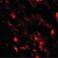
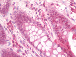
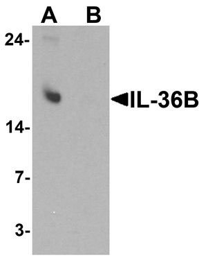
| WB, IHC-P, IF, E |
|---|---|
| Primary Accession | Q9NZH7 |
| Reactivity | Human |
| Host | Rabbit |
| Clonality | Polyclonal |
| Calculated MW | 19kDa |
| Dilution | IF (20 µg/ml), IHC-P (10 µg/ml), WB (1-2 µg/ml) |
| Gene ID | 27177 |
|---|---|
| Other Names | Interleukin-36 beta, FIL1 eta, Interleukin-1 eta, IL-1 eta, Interleukin-1 family member 8, IL-1F8, Interleukin-1 homolog 2, IL-1H2, IL36B, IL1F8, IL1H2 |
| Target/Specificity | IL-36B antibody is human specific. At least two isoforms of IL-36B are known to exist; this antibody will only detect the longer isoform. IL-36B antibody will not cross-react with IL-36A or IL-36G. |
| Reconstitution & Storage | Long term: -20°C; Short term: +4°C. Avoid repeat freeze-thaw cycles. |
| Precautions | IL36B Antibody (C-Terminus) is for research use only and not for use in diagnostic or therapeutic procedures. |
| Name | IL36B (HGNC:15564) |
|---|---|
| Synonyms | IL1F8, IL1H2 |
| Function | Cytokine that binds to and signals through the IL1RL2/IL-36R receptor which in turn activates NF-kappa-B and MAPK signaling pathways in target cells linked to a pro-inflammatory response. Part of the IL- 36 signaling system that is thought to be present in epithelial barriers and to take part in local inflammatory response; similar to the IL-1 system with which it shares the coreceptor IL1RAP. Stimulates production of interleukin-6 and interleukin-8 in synovial fibrobasts, articular chondrocytes and mature adipocytes. Induces expression of a number of antimicrobial peptides including beta-defensins 4 and 103 as well as a number of matrix metalloproteases. Seems to be involved in skin inflammatory response by acting on keratinocytes, dendritic cells and indirectly on T-cells to drive tissue infiltration, cell maturation and cell proliferation. In cultured keratinocytes induces the expression of macrophage, T-cell, and neutrophil chemokines, such as CCL3, CCL4, CCL5, CCL2, CCL17, CCL22, CL20, CCL5, CCL2, CCL17, CCL22, CXCL8, CCL20 and CXCL1, and the production of pro-inflammatory cytokines such as TNF-alpha, IL-8 and IL-6. |
| Cellular Location | Cytoplasm. Secreted. Note=The secretion is dependent on protein unfolding and facilitated by the cargo receptor TMED10; it results in protein translocation from the cytoplasm into the ERGIC (endoplasmic reticulum-Golgi intermediate compartment) followed by vesicle entry and secretion. |
| Tissue Location | Expression at low levels in tonsil, bone marrow, heart, placenta, lung, testis and colon but not in any hematopoietic cell lines. Not detected in adipose tissue. Expressed at higher levels in psoriatic plaques than in symptomless psoriatic skin or healthy control skin. Increased levels are not detected in inflamed joint tissue. |

Thousands of laboratories across the world have published research that depended on the performance of antibodies from Abcepta to advance their research. Check out links to articles that cite our products in major peer-reviewed journals, organized by research category.
info@abcepta.com, and receive a free "I Love Antibodies" mug.

Provided below are standard protocols that you may find useful for product applications.
Background
Cytokine that binds to and signals through the IL1RL2/IL-36R receptor which in turn activates NF-kappa-B and MAPK signaling pathways in target cells linked to a pro-inflammatory response. Part of the IL-36 signaling system that is thought to be present in epithelial barriers and to take part in local inflammatory response; similar to the IL-1 system with which it shares the coreceptor IL1RAP. Stimulates production of interleukin-6 and interleukin-8 in synovial fibrobasts, articular chondrocytes and mature adipocytes. Induces expression of a number of antimicrobial peptides including beta-defensins 4 and 103 as well as a number of matrix metalloproteases. Seems to be involved in skin inflammatory response by acting on keratinocytes, dendritic cells and indirectly on T cells to drive tissue infiltration, cell maturation and cell proliferation. In cultured keratinocytes induces the expression of macrophage, T cell, and neutrophil chemokines, such as CCL3, CCL4, CCL5, CCL2, CCL17, CCL22, CL20, CCL5, CCL2, CCL17, CCL22, CXCL8, CCL20 and CXCL1, and the production of proinflammatory cytokines such as TNF-alpha, IL- 8 and IL-6.
References
Kumar S.,et al.J. Biol. Chem. 275:10308-10314(2000).
Smith D.E.,et al.J. Biol. Chem. 275:1169-1175(2000).
Nicklin M.J.H.,et al.Genomics 79:718-725(2002).
Hillier L.W.,et al.Nature 434:724-731(2005).
Mural R.J.,et al.Submitted (JUL-2005) to the EMBL/GenBank/DDBJ databases.
If you have used an Abcepta product and would like to share how it has performed, please click on the "Submit Review" button and provide the requested information. Our staff will examine and post your review and contact you if needed.
If you have any additional inquiries please email technical services at tech@abcepta.com.
Ordering Information
Other Products
Shipping Information
