


 Foundational characteristics of cancer include proliferation, angiogenesis, migration, evasion of apoptosis, and cellular immortality. Find key markers for these cellular processes and antibodies to detect them.
 The SUMOplot™ Analysis Program predicts and scores sumoylation sites in your protein. SUMOylation is a post-translational modification involved in various cellular processes, such as nuclear-cytosolic transport, transcriptional regulation, apoptosis, protein stability, response to stress, and progression through the cell cycle.
 The Autophagy Receptor Motif Plotter predicts and scores autophagy receptor binding sites in your protein. Identifying proteins connected to this pathway is critical to understanding the role of autophagy in physiological as well as pathological processes such as development, differentiation, neurodegenerative diseases, stress, infection, and cancer.
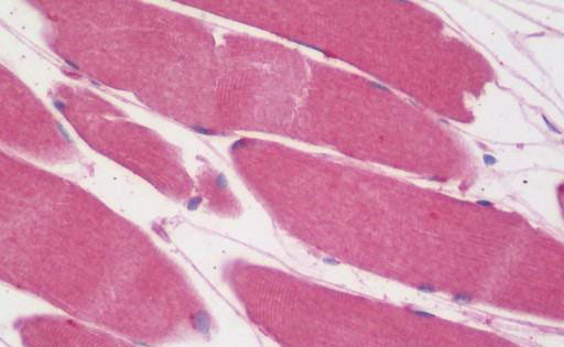
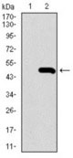
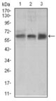
ALK3 / BMPR1A Antibody (aa179-378, clone 4B7B2)
Mouse Monoclonal Antibody
- SPECIFICATION
- CITATIONS
- PROTOCOLS
- BACKGROUND

Application
| WB, IHC-P, ICC, E, FC |
|---|---|
| Primary Accession | P36894 |
| Reactivity | Human, Mouse |
| Host | Mouse |
| Clonality | Monoclonal |
| Clone Names | 4B7B2 |
| Calculated MW | 60kDa |
| Dilution | ELISA (1:10000), Flo (1:200-1:400), ICC (1:200-1:1000), IHC-P (1:50), WB (1:500-1:2000) |
| Gene ID | 657 |
|---|---|
| Other Names | Bone morphogenetic protein receptor type-1A, BMP type-1A receptor, BMPR-1A, 2.7.11.30, Activin receptor-like kinase 3, ALK-3, Serine/threonine-protein kinase receptor R5, SKR5, CD292, BMPR1A, ACVRLK3, ALK3 |
| Target/Specificity | Human ALK3 / BMPR1A |
| Reconstitution & Storage | Short term +4°C; Long term -20°C |
| Precautions | ALK3 / BMPR1A Antibody (aa179-378, clone 4B7B2) is for research use only and not for use in diagnostic or therapeutic procedures. |
| Name | BMPR1A |
|---|---|
| Synonyms | ACVRLK3, ALK3 |
| Function | On ligand binding, forms a receptor complex consisting of two type II and two type I transmembrane serine/threonine kinases. Type II receptors phosphorylate and activate type I receptors which autophosphorylate, then bind and activate SMAD transcriptional regulators. Receptor for BMP2, BMP4, GDF5 and GDF6. Positively regulates chondrocyte differentiation through GDF5 interaction. Mediates induction of adipogenesis by GDF6. May promote the expression of HAMP, potentially via its interaction with BMP2 (By similarity). |
| Cellular Location | Cell membrane; Single-pass type I membrane protein. Cell surface {ECO:0000250|UniProtKB:P36895} |
| Tissue Location | Highly expressed in skeletal muscle. |
| Volume | 50 µl |

Thousands of laboratories across the world have published research that depended on the performance of antibodies from Abcepta to advance their research. Check out links to articles that cite our products in major peer-reviewed journals, organized by research category.
info@abcepta.com, and receive a free "I Love Antibodies" mug.

Provided below are standard protocols that you may find useful for product applications.
Background
On ligand binding, forms a receptor complex consisting of two type II and two type I transmembrane serine/threonine kinases. Type II receptors phosphorylate and activate type I receptors which autophosphorylate, then bind and activate SMAD transcriptional regulators. Receptor for BMP-2 and BMP-4.
References
ten Dijke P.,et al.Oncogene 8:2879-2887(1993).
Ota T.,et al.Nat. Genet. 36:40-45(2004).
Mahlawat P.,et al.Biochemistry 51:6328-6341(2012).
Kirsch T.,et al.Nat. Struct. Biol. 7:492-496(2000).
Howe J.R.,et al.Nat. Genet. 28:184-187(2001).
If you have used an Abcepta product and would like to share how it has performed, please click on the "Submit Review" button and provide the requested information. Our staff will examine and post your review and contact you if needed.
If you have any additional inquiries please email technical services at tech@abcepta.com.
Ordering Information
Other Products
Shipping Information
