


 Foundational characteristics of cancer include proliferation, angiogenesis, migration, evasion of apoptosis, and cellular immortality. Find key markers for these cellular processes and antibodies to detect them.
 The SUMOplot™ Analysis Program predicts and scores sumoylation sites in your protein. SUMOylation is a post-translational modification involved in various cellular processes, such as nuclear-cytosolic transport, transcriptional regulation, apoptosis, protein stability, response to stress, and progression through the cell cycle.
 The Autophagy Receptor Motif Plotter predicts and scores autophagy receptor binding sites in your protein. Identifying proteins connected to this pathway is critical to understanding the role of autophagy in physiological as well as pathological processes such as development, differentiation, neurodegenerative diseases, stress, infection, and cancer.
ODZ3 Antibody (N-Terminus)
Rabbit Polyclonal Antibody
- SPECIFICATION
- CITATIONS
- PROTOCOLS
- BACKGROUND

Application
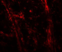
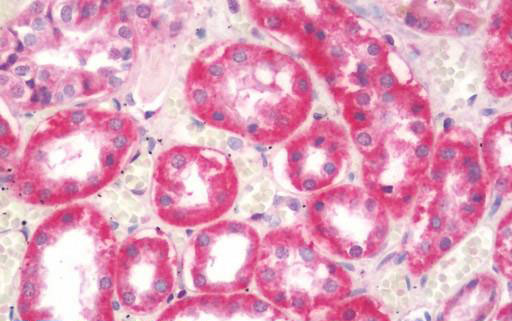
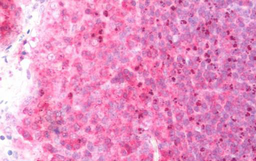
| WB, IHC-P, IF, E |
|---|---|
| Primary Accession | Q9P273 |
| Reactivity | Human, Mouse, Rat |
| Host | Rabbit |
| Clonality | Polyclonal |
| Calculated MW | 301kDa |
| Dilution | IHC-P (10 µg/ml), WB (1-2 µg/ml), |
| Gene ID | 55714 |
|---|---|
| Other Names | Teneurin-3, Ten-3, Protein Odd Oz/ten-m homolog 3, Tenascin-M3, Ten-m3, Teneurin transmembrane protein 3, TENM3, KIAA1455, ODZ3, TNM3 |
| Target/Specificity | TENM3 antibody is human, mouse and rat reactive. TENM3 antibody is predicted to not cross-react with other members of the TENM family. |
| Reconstitution & Storage | Long term: -20°C; Short term: +4°C. Avoid repeat freeze-thaw cycles. |
| Precautions | ODZ3 Antibody (N-Terminus) is for research use only and not for use in diagnostic or therapeutic procedures. |
| Name | TENM3 (HGNC:29944) |
|---|---|
| Function | Involved in neural development by regulating the establishment of proper connectivity within the nervous system. Acts in both pre- and postsynaptic neurons in the hippocampus to control the assembly of a precise topographic projection: required in both CA1 and subicular neurons for the precise targeting of proximal CA1 axons to distal subiculum, probably by promoting homophilic cell adhesion. Required for proper dendrite morphogenesis and axon targeting in the vertebrate visual system, thereby playing a key role in the development of the visual pathway. Regulates the formation in ipsilateral retinal mapping to both the dorsal lateral geniculate nucleus (dLGN) and the superior colliculus (SC). May also be involved in the differentiation of the fibroblast-like cells in the superficial layer of mandibular condylar cartilage into chondrocytes. |
| Cellular Location | Cell membrane {ECO:0000250|UniProtKB:Q9WTS6}; Single-pass membrane protein {ECO:0000250|UniProtKB:Q9WTS6}. Cell projection, axon {ECO:0000250|UniProtKB:Q9WTS6} |
| Tissue Location | Expressed in adult and fetal brain, slightly lower levels in testis and ovary, and intermediate levels in all other peripheral tissues examined. Not expressed in spleen or liver Expression was high in brain, with highest levels in amygdala and caudate nucleus, followed by thalamus and subthalamic nucleus |

Thousands of laboratories across the world have published research that depended on the performance of antibodies from Abcepta to advance their research. Check out links to articles that cite our products in major peer-reviewed journals, organized by research category.
info@abcepta.com, and receive a free "I Love Antibodies" mug.

Provided below are standard protocols that you may find useful for product applications.
Background
Involved in neural development, regulating the establishment of proper connectivity within the nervous system. Promotes axon guidance and homophilic cell adhesion. Plays a role in the development of the visual pathway; regulates the formation in ipsilateral retinal mapping to both the dorsal lateral geniculate nucleus (dLGN) and the superior colliculus (SC). May be involved in the differentiation of the fibroblast-like cells in the superficial layer of mandibular condylar cartilage into chondrocytes. May function as a cellular signal transducer (By similarity).
References
Hillier L.W.,et al.Nature 434:724-731(2005).
Ben-Zur T.,et al.Dev. Biol. 217:107-120(2000).
Nagase T.,et al.DNA Res. 7:143-150(2000).
Ota T.,et al.Nat. Genet. 36:40-45(2004).
Steckler N.K.,et al.Submitted (AUG-2004) to the EMBL/GenBank/DDBJ databases.
If you have used an Abcepta product and would like to share how it has performed, please click on the "Submit Review" button and provide the requested information. Our staff will examine and post your review and contact you if needed.
If you have any additional inquiries please email technical services at tech@abcepta.com.
Ordering Information
Other Products
Shipping Information
