


 Foundational characteristics of cancer include proliferation, angiogenesis, migration, evasion of apoptosis, and cellular immortality. Find key markers for these cellular processes and antibodies to detect them.
 The SUMOplot™ Analysis Program predicts and scores sumoylation sites in your protein. SUMOylation is a post-translational modification involved in various cellular processes, such as nuclear-cytosolic transport, transcriptional regulation, apoptosis, protein stability, response to stress, and progression through the cell cycle.
 The Autophagy Receptor Motif Plotter predicts and scores autophagy receptor binding sites in your protein. Identifying proteins connected to this pathway is critical to understanding the role of autophagy in physiological as well as pathological processes such as development, differentiation, neurodegenerative diseases, stress, infection, and cancer.
JAKMIP1 Antibody (Internal)
Rabbit Polyclonal Antibody
- SPECIFICATION
- CITATIONS
- PROTOCOLS
- BACKGROUND

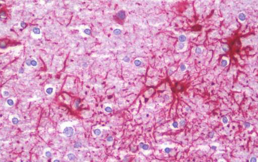
Application
| WB, IHC-P, E |
|---|---|
| Primary Accession | Q96N16 |
| Other Accession | 152789 |
| Reactivity | Human, Mouse, Rat |
| Host | Rabbit |
| Clonality | Polyclonal |
| Isotype | IgG |
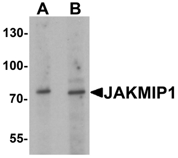
| Calculated MW | 73209 Da |
| Dilution | IHC-P (10 µg/ml), WB (1 - 2 µg/ml), |
| Gene ID | 152789 |
|---|---|
| Other Names | JAKMIP1, JAMIP1, Gababrbp, Marlin-1, MARLIN1 |
| Target/Specificity | Multiple isoforms of JAKMIP1 are known to exist. JAKMIP1 antibody is predicted to not cross-react with JAKMIP2. |
| Reconstitution & Storage | PBS, 0.02% sodium azide. Long term: -20°C; Short term: +4°C. Avoid repeat freeze-thaw cycles. |
| Precautions | JAKMIP1 Antibody (Internal) is for research use only and not for use in diagnostic or therapeutic procedures. |
| Name | JAKMIP1 |
|---|---|
| Synonyms | GABABRBP, JAMIP1, MARLIN1 |
| Function | Associates with microtubules and may play a role in the microtubule-dependent transport of the GABA-B receptor. May play a role in JAK1 signaling and regulate microtubule cytoskeleton rearrangements. |
| Cellular Location | Cytoplasm, cytoskeleton. Membrane; Peripheral membrane protein. Note=Colocalizes with the microtubule network Localizes to the cell body and neurites of hippocampal neurons where it accumulates in granules. Localizes to the tail and to a lower extent to the head of sperm cells |
| Tissue Location | Predominantly expressed in neural tissues and lymphoid cells (at protein level). Isoform 2, isoform 3 and isoform 4 are specifically expressed in brain and retina. Isoform 1 and isoform 5 are also detected in liver, lung and skeletal muscle. Also detected in testis and to a lower extent spleen and intestine |

Thousands of laboratories across the world have published research that depended on the performance of antibodies from Abcepta to advance their research. Check out links to articles that cite our products in major peer-reviewed journals, organized by research category.
info@abcepta.com, and receive a free "I Love Antibodies" mug.

Provided below are standard protocols that you may find useful for product applications.
Background
Associates with microtubules and may play a role in the microtubule-dependent transport of the GABA-B receptor. May play a role in JAK1 signaling and regulate microtubule cytoskeleton rearrangements.
References
Couve A.,et al.J. Biol. Chem. 279:13934-13943(2004).
Costa V.,et al.Gene 402:1-8(2007).
Ota T.,et al.Nat. Genet. 36:40-45(2004).
Hillier L.W.,et al.Nature 434:724-731(2005).
Mural R.J.,et al.Submitted (SEP-2005) to the EMBL/GenBank/DDBJ databases.
If you have used an Abcepta product and would like to share how it has performed, please click on the "Submit Review" button and provide the requested information. Our staff will examine and post your review and contact you if needed.
If you have any additional inquiries please email technical services at tech@abcepta.com.

Ordering Information
Other Products
Shipping Information
