


 Foundational characteristics of cancer include proliferation, angiogenesis, migration, evasion of apoptosis, and cellular immortality. Find key markers for these cellular processes and antibodies to detect them.
 The SUMOplot™ Analysis Program predicts and scores sumoylation sites in your protein. SUMOylation is a post-translational modification involved in various cellular processes, such as nuclear-cytosolic transport, transcriptional regulation, apoptosis, protein stability, response to stress, and progression through the cell cycle.
 The Autophagy Receptor Motif Plotter predicts and scores autophagy receptor binding sites in your protein. Identifying proteins connected to this pathway is critical to understanding the role of autophagy in physiological as well as pathological processes such as development, differentiation, neurodegenerative diseases, stress, infection, and cancer.
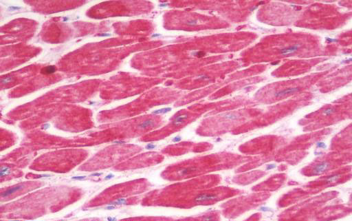
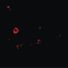
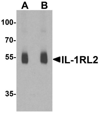
IL1RL2 Antibody (Internal)
Rabbit Polyclonal Antibody
- SPECIFICATION
- CITATIONS
- PROTOCOLS
- BACKGROUND

Application
| WB, IHC-P, IF, E |
|---|---|
| Primary Accession | Q9HB29 |
| Other Accession | 8808 |
| Reactivity | Human, Mouse, Rat |
| Host | Rabbit |
| Clonality | Polyclonal |
| Isotype | IgG |
| Calculated MW | 65405 Da |
| Dilution | IF (20 µg/ml), IHC-P (10 µg/ml), WB (1 - 2 µg/ml), |
| Gene ID | 8808 |
|---|---|
| Other Names | IL1RL2, IL-1Rrp2, IL1R-rp2, IL1RRP2, Interleukin 1 receptor-like 2, Interleukin-1 receptor-like 2, IL-36R |
| Target/Specificity | IL-1RL2 antibody is human, mouse and rat reactive. At least three isoforms of IL-1RL2 are known to exist; this antibody will detect all three isoforms. IL-1RL2 antibody is predicted to not cross-react with IL-1R or IL-1RL1. |
| Reconstitution & Storage | PBS, 0.02% sodium azide. Long term: -20°C; Short term: +4°C. Avoid repeat freeze-thaw cycles. |
| Precautions | IL1RL2 Antibody (Internal) is for research use only and not for use in diagnostic or therapeutic procedures. |
| Name | IL1RL2 |
|---|---|
| Synonyms | IL1RRP2 |
| Function | Receptor for interleukin-36 (IL36A, IL36B and IL36G). After binding to interleukin-36 associates with the coreceptor IL1RAP to form the interleukin-36 receptor complex which mediates interleukin-36- dependent activation of NF-kappa-B, MAPK and other pathways (By similarity). The IL-36 signaling system is thought to be present in epithelial barriers and to take part in local inflammatory response; it is similar to the IL-1 system. Seems to be involved in skin inflammatory response by induction of the IL-23/IL-17/IL-22 pathway. |
| Cellular Location | Membrane; Single-pass type I membrane protein |
| Tissue Location | Expressed in synovial fibroblasts and articular chondrocytes. Expressed in keratinocytes and monocyte-derived dendritic cells. Expressed in monocytes and myeloid dendritic cells; at protein level. |

Thousands of laboratories across the world have published research that depended on the performance of antibodies from Abcepta to advance their research. Check out links to articles that cite our products in major peer-reviewed journals, organized by research category.
info@abcepta.com, and receive a free "I Love Antibodies" mug.

Provided below are standard protocols that you may find useful for product applications.
Background
Receptor for interleukin-36 (IL36A, IL36B and IL36G). After binding to interleukin-36 associates with the coreceptor IL1RAP to form the interleukin-36 receptor complex which mediates interleukin-36-dependent activation of NF-kappa-B, MAPK and other pathways (By similarity). The IL-36 signaling system is thought to be present in epithelial barriers and to take part in local inflammatory response; it is similar to the IL-1 system. Seems to be involved in skin inflammatory response by induction of the IL- 23/IL-17/IL-22 pathway.
References
Born T.L.,et al.J. Biol. Chem. 275:29946-29954(2000).
Lovenberg T.W.,et al.J. Neuroimmunol. 70:113-122(1996).
Hillier L.W.,et al.Nature 434:724-731(2005).
Debets R.,et al.J. Immunol. 167:1440-1446(2001).
Magne D.,et al.Arthritis Res. Ther. 8:R80-R80(2006).
If you have used an Abcepta product and would like to share how it has performed, please click on the "Submit Review" button and provide the requested information. Our staff will examine and post your review and contact you if needed.
If you have any additional inquiries please email technical services at tech@abcepta.com.
Ordering Information
Other Products
Shipping Information
